救急一直線 特別ブログ Happy保存の法則 ー United in the World for Us ー

HP「救急一直線〜Happy保存の法則〜」は,2002年に開始され,現在はブログとして継続されています。

DIPNECH 2015年 Diffuse Idiopathic Pulmonary Neuroendocrine Cell Hyperplasia of the Lung

2015年09月21日 02時50分53秒 | 総説紹介
Diffuse Idiopathic Pulmonary Neuroendocrine Cell Hyperplasia of the Lung (DIPNECH): Current Best Evidence.
Wirtschafter E1, Walts AE, Liu ST, Marchevsky AM.
Lung. 2015 Jun 24.

Diffuse idiopathic pulmonary neuroendocrine cell hyperplasia (DIPNECH) is recognized as a preneoplastic condition by the World Health Organization. We reviewed our experience with 30 patients and performed a systematic review of the English literature to collect best evidence on the clinical features and disease course in 169 additional patients. Some patients presented with one or more carcinoid tumors associated with multiple small pulmonary nodules on imaging studies and showed DIPNECH as a somewhat unexpected pathologic finding. Others presented with multiple small pulmonary nodules that raised suspicion of metastatic disease on imaging. A third subset was presented with previously unexplained respiratory symptoms. In most patients, DIPNECH was associated with a good prognosis, with chronological progression into a subsequent carcinoid tumor noted in only one patient and death attributed directly to DIPNECH in only two patients. There is no best evidence to support the use of octreotide, steroids, or bronchodilators in DIPNECH patients.

http://pubs.rsna.org/doi/full/10.1148/rg.336135506

DIPNECH: when to suggest this diagnosis on CT.
Chassagnon G, Favelle O, Marchand-Adam S, De Muret A, Revel MP.
Clin Radiol. 2015;70:317-25.

(2014) Sarcoid-Like Reaction in Diffuse Idiopathic Pulmonary Neuroendocrine Cell Hyperplasia. American Journal of Respiratory and Critical Care Medicine 190:10, e62-e63

(2012) mTOR/p70S6K in Diffuse Idiopathic Pulmonary Neuroendocrine Cell Hyperplasia. American Journal of Respiratory and Critical Care Medicine 185:3, 341-342

(2011) Diffuse Idiopathic Pulmonary Neuroendocrine Cell Hyperplasia:A Systematic Overview
Am J Respir Crit Care Med184:8–16

※ 当科の管理するoncological ICUの肺傷害の管理として,念頭に置いています。

  • X
  • Facebookでシェアする
  • はてなブックマークに追加する
  • LINEでシェアする

総説 虚血性指肢壊疽

2015年08月18日 02時41分16秒 | 総説紹介
Ischemic Limb Gangrene with Pulses
Warkentin TE.
N Engl J Med. 2015 Aug 13;373(7):642-55.


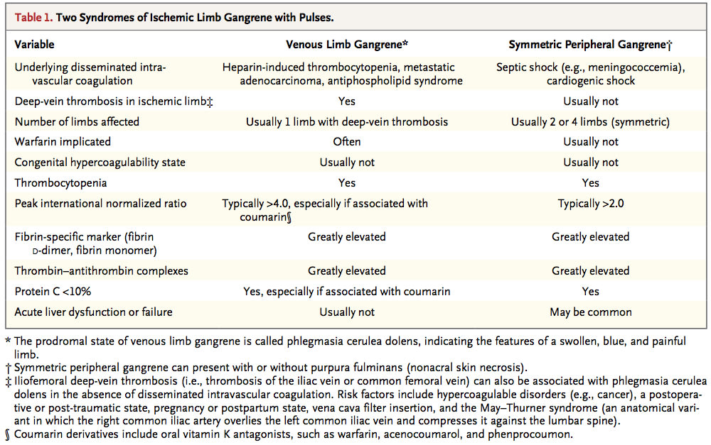
There are two distinct syndromes of microthrombosis-associated ischemic limb injury (Table 1). Venous limb gangrene can complicate thrombocytopenic disor- ders that are strongly associated with deep-vein thrombosis (e.g., cancer-associat- ed disseminated intravascular coagulation and heparin-induced thrombocytope- nia). In these conditions, microthrombosis occurs in the same limb with acute large-vein thrombosis, resulting in acral (distal-extremity) ischemic necrosis. Usually, only one limb is affected. The potentially reversible, prodromal state of limb-threatening ischemia is phlegmasia cerulea dolens, indicating the respective features of a swollen, blue (ischemic), and painful limb (Fig. 1A).
 In contrast, two and sometimes all four limbs are affected in symmetric pe- ripheral gangrene, also featuring acral limb ischemic necrosis but usually without deep-vein thrombosis (Fig. 1B). The limb necrosis is often strikingly symmetric; lower limbs are most often affected, with additional involvement of fingers or hands in approximately one third of patients. When there is additional or pre- dominant nonacral tissue necrosis, the term purpura fulminans is applicable. Patients are usually critically ill, with cardiogenic or septic shock. In 1904, Barraud discussed limb gangrene as a complication of acute infection, a complication that continues to occur today. The two syndromes have common pathophysiological features of micro- thrombosis associated with a disturbed procoagulant–anticoagulant balance (Fig. 1C).
 The concept that venous limb gangrene and sym- metric peripheral gangrene are usually associated with microvascular thrombosis with underlying disseminated intravascular coagulation provides a framework for a rational approach to diagnos- ing and treating these diverse and potentially devastating disorders. Prevention and treatment of venous gangrene requires correction of abnor- malities associated with the use of vitamin K an- tagonists and aggressive anticoagulation, whereas treatment of symmetric peripheral gangrene (with or without purpura fulminans) theoretically in- volves heparin-based anticoagulation and the sub- stitution of natural anticoagulants.


  • X
  • Facebookでシェアする
  • はてなブックマークに追加する
  • LINEでシェアする

総説 糖尿病による電解質異常のメカニズム

2015年08月12日 02時45分18秒 | 総説紹介
Electrolyte and Acid-Base Disturbances in Patients with Diabetes Mellitus.
Palmer BF, Clegg DJ.
N Engl J Med. 2015;373:548-59.
Dr. Palmer at the Department of Internal Medicine, University of Texas Southwest- ern Medical Center, 5323 Harry Hines Blvd., Dallas, TX 75390, or at biff.palmer@ utsouthwestern.edu.


The prevalence of diabetes is increasing rapidly, and type 2 diabetes now accounts for 20 to 50% of cases of new-onset diabetes in young people.1 Electrolyte disturbances are common in patients with diabetes and may be the result of an altered distribution of electrolytes related to hyperglycemia-induced osmotic fluid shifts or of total-body deficits brought about by osmotic diuresis. Complications from end-organ injury and the therapies used in the management of diabetes may also contribute to electrolyte disturbances. In this review, we highlight the ways in which specific electrolytes may be influenced by dysregulation in glucose homeostasis.
The dysregulation of glucose homeostasis leads to many direct and indirect effects on electrolyte and acid–base balance. Since the high prevalence of diabetes ensures that clinicians in virtually every medical specialty will interact with these patients, familiarity with related electrolyte abnormalities is important.


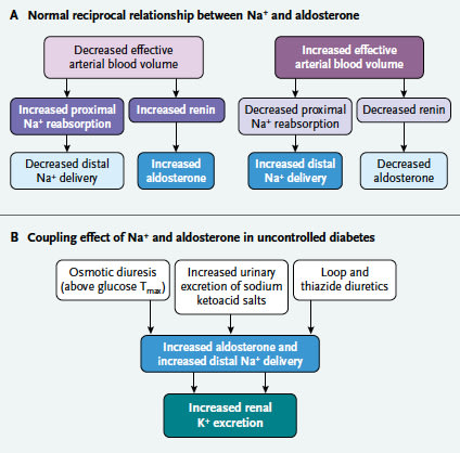
Figure 1. Volume Regulation in Persons with and without Diabetes.
In the regulation of effective arterial blood volume (Panel A), there is a
balanced, reciprocal relationship between the delivery of sodium to the distal nephron and the circulating level of aldosterone that serves to main- tain potassium balance. In patients with uncontrolled diabetes (Panel B), the osmotic diuretic effect of glucose (glucose Tmax denotes the maximum rate of the reabsorption of glucose in the proximal tubule) and the excre- tion of sodium ketoacid salts cause an increase in the delivery of sodium to the distal nephron. At the same time, mineralocorticoid activity is in- creased in response to volume depletion. The coupling of the increased delivery of sodium with the increased mineralocorticoid activity results in renal potassium wasting and total-body depletion. The use of loop or thia- zide diuretics also contributes to renal potassium wasting by means of this coupling effect. In addition, high flow rates in the distal nephron lower the luminal potassium concentration, providing a more favorable gradient for the diffusion of potassium into the luminal fluid. High flow in the distal nephron also activates potassium secretion by means of the calcium- activated potassium channel (or the maxi-K+channel).


Figure 2. Phases of Metabolic Acidosis in Patients with Diabetes.
In the early phase of ketoacidosis, when the volume of extracellular fluid (ECF) is close to normal, the ketoacid anions produced will
be rapidly excreted by the kidney as sodium and potassium salts. The urinary loss of ketone salts leads to the contraction of the volume of ECF and signals the renal retention of dietary sodium chloride. The proton of the ketoacid reacts with bicarbonate to generate water and carbon dioxide, which are expired through the lungs. The net effect is the development of a hyperchloremic normal-gap acidosis. This process has been referred to as an indirect loss of sodium bicarbonate.7 As the ketogenic process becomes more accelerated and as volume depletion becomes more severe, a larger proportion of the generated ketoacid salts are retained within the body, thus in- creasing the anion gap. At this point, glomerular filtration rate (GFR) is typically reduced and a patient requires treatment and admis- sion to a hospital. During the recovery phase, the anion-gap metabolic acidosis is transformed once again into a hyperchloremic, nor- mal anion-gap acidosis. Treatment leads to the termination of ketoacid production. As the ECF volume is restored, there is increased renal excretion of the sodium salts of the ketoacid anions. The indirect loss of bicarbonate, combined with the retention of adminis- tered sodium chloride, accounts for the redevelopment of the hyperchloremic, normal-gap acidosis. In addition, the potassium and sodium administered in solutions containing sodium chloride and potassium chloride enter into cells in exchange for hydrogen ions. The net effect is the infusion of hydrogen chloride into the extracellular fluid. The normalization of the acid–base balance is accom- plished over a period of several days as the bicarbonate deficit is corrected as bicarbonate is regenerated by the kidney.


Figure 3. Pathogenesis of the Electrolyte Pattern in Type 4 Renal Tubular Acidosis.
In normal circumstances, the reabsorption of sodium in the collecting duct, driven by aldosterone, generates
negative potential in the lumen, which serves as a driving force for the secretion of potassium by the principal cell and of hydrogen ions by the α-intercalated cell. Impaired sodium reabsorption in the principal cell ― caused by either hyporeninemic hypoaldosteronism or impairment in the function of the collecting duct ― leads to a decrease in luminal electronegativity. This decrease impairs secretion of potassium and of hydrogen ions, contributing to hy- perkalemia and metabolic acidosis. The hyperkalemia further impairs acidification by decreasing the amount of ammonium available to act as a urinary buffer. First, hyperkalemia decreases the production of ammonium in the proximal tubule. The precise mechanism by which this occurs is not currently known, but it may involve the entry of potassium into cells in exchange for protons, which would raise the intracellular pH. Second, the transport of ammonium in the thick ascending limb is inhibited by the large increase in the concentration of potassium in the lumen, which effectively competes with ammonium for transport on the sodium–potassium–chloride cotransporter. Ammonium normally exits the basolateral surface of the cell through sodium–proton exchanger 4 (NHE4). The net excretion of acid decreases as a result of the limited availability of a buffer combined with a decreased capacity for the secretion of hydrogen ions. The urinary osmolal gap is not increased, which indicates that there is little or no excretion of ammonium in the urine. Patients in whom type 4 renal tubular acidosis is caused by a defect in mineralo- corticoid activity typically have a urinary pH of less than 5.5, reflecting a more severe defect in the availability of ammonium than in the secretion of hydrogen ions. In patients with structural damage, the secretion of hydrogen ions is impaired throughout the collecting duct (both cortical and medullary segments) such that the urinary pH may be more alkaline than it is in patients who have impaired mineralocorticoid activity alone.

  • X
  • Facebookでシェアする
  • はてなブックマークに追加する
  • LINEでシェアする