画像診断
日本語の語順からすればあくまで「画像を用いた診断」のような気がしますが、対応する英語としてはdiagnostic imagingや単なるimagingが多い印象で、それを直訳すれば「診断のための撮像」や「イメージング」となって、意味が異なるような気がしますがどうなんでしょうか?
つまり、「画像診断」と聞くとあくまで重点、目的、動作は「診断」に感じるのですが、英語のdiagnostic imagingは重点、目的、動作はimaging、「撮像」ではないかと感じます。
diagnostic imaging:診断撮像、診断イメージングと訳したらどうでしょう?
まあ今更無理でしょうが。
(ただし、もし本当に「画像を診断する」であればimage is diagnosedで、その意味での「画像診断装置」ならimage diagnosing deviceやimage diagnosis device等で良いのではと思います。)
「多くの画像診断医は目の前の画像をみて,肝細胞癌の有無,門脈圧亢進症による合併症など,ある程度決まった手順で診断し」(画像診断 Vol.42 No.1 2022年1月号)
(上記の専門誌の英語タイトルはJapanese Journal of Imaging Diagnosis)
US8937157
In contrast, Fc variants with decreased FcRn binding affinity are expected to have shorter half-lives,
対照的に、FcRn結合親和性が低下したFc変異体はより短い半減期を有すると予想され、
and such molecules are also useful, for example, for administration to a mammal where a shortened circulation time may be advantageous, e.g. for in vivo diagnostic imaging or in situations where the starting polypeptide has toxic side effects when present in the circulation for prolonged periods.
そのような分子も、短縮した循環時間が有用と考えられるような哺乳動物に対する投与のために、例えばインビボ画像診断(この時点では「診断」というより「撮像」では?)のために、または出発ポリペプチドが循環血中に長期にわたって存在すると有害副作用を有する状況においては有用である。
EP3972596
This latter could occur, for example and not limitation, from any one or more of the following: demonstration of NASH resolution and/or from an improvement in the fibrosis score based on liver biopsy;
この後者は、たとえば、限定はせず、以下の事項、すなわち、NASH解消の確証および/または肝生検に基づく線維化スコアの改善;
lower incidence of progression to cirrhosis, hepatocellular carcinoma, and/or other liver related outcomes;
硬変、肝細胞癌、および/または肝臓に関連した他の結果への進行のより低い出現率;
a reduction or improvement of the level of serum or imaging based markers of nonalcoholic steatohepatitis activity;
非アルコール性脂肪性肝炎活性の血清または画像診断に基づくマーカーのレベルの低下または改善;
reduction or improvement of nonalcoholic steatohepatitis disease activity; or reduction in the medical consequences of nonalcoholic steatohepatitis.非アルコール性脂肪性肝炎疾患活性の低下または改善;非アルコール性脂肪性肝炎の医学的帰結の減少のいずれか1つまたは複数により生じる場合がある。
EP3116311
Assays can include functional evaluation of the transplanted tissue matrix or imaging techniques (e.g., computed tomography (CT),
外科医は、磁気共鳴画像診断(MRI)システム、コンピュータ断層撮影(CT)システム、ultrasound, or magnetic resonance imaging (e.g., contrast-enhanced MRI)).
アッセイは、移植された組織マトリックスの機能的評価、または撮像技術(例えば、コンピュータ断層撮影(CT)、超音波、もしくは磁気共鳴画像診断(例えば、コントラスト強調MRI))を含むことができる。
US10758194
A surgeon
外科医は、
can perform the procedure on the subject with images of the subject that can be acquired using imaging systems such as
a magnetic resonance imaging (MRI) system, computed tomography (CT) system,
磁気共鳴画像診断(MRI)システム、コンピュータ断層撮影(CT)システム、
fluoroscopy (e.g. C-Arm imaging systems), or other appropriate imaging systems.
蛍光透視法(例えば、Cアーム画像診断(*いや撮像、イメージングでは?)システム)、または他の適切な画像診断システム
などの画像診断システムを使用して取得することができる対象の画像を用いて、対象に対して処置を実行することができる。
US2022175306
[0082] The controller 16 may also be configured to receive inputs related to the targeted physiological outcomes as an input to the selection of the modulation parameters.
【0057】
コントローラ16はまた、調節パラメータの選択に対する入力として標的生理学的結果に関連する入力を受け取るように構成されていてもよい。
For example, when an imaging modality is used to assess a tissue characteristic, the controller 16 may be configured to receive a calculated index or parameter of the characteristic.
例えば、画像診断法を使用して組織特性を評価する時には、コントローラ16は特性の計算された指標又はパラメータを受け取るように構成されていてもよい。
US2016303284
The location of the tip of the guide needle or the location of the tip of the injection needle can be verified using imaging technology, e.g., fluoroscopy, magnetic resonance imaging, computed tomography, or any other similar technology well known in the art.
ガイド針の先端の位置、または注入針の先端の位置は、例えば蛍光透視法、磁気共鳴画像法、コンピューター断層切断法、または当技術分野において既知で任意である他の同様の技術などの画像診断を用いて、確認することが可能である。
US10010546
Diagnosis and monitoring of the diseases is
これらの疾患の診断およびモニタリングは、
performed according to methods and diagnostic tests routinely practiced in the art, including blood tests, colonoscopy, flexible sigmoidoscopy, barium enema, CT scan, MRI, endoscopy, and small intestine imaging.
血液検査、結腸内視鏡検査、軟性S状結腸鏡検査、バリウム注腸、CTスキャン、MRI、内視鏡検査、および小腸画像診断を含む、当該技術分野で慣例的に実施される方法および診断テストに従って実行される。
US2020197396
Patients were screened prior to administration of a combination therapy described herein and again at each subsequent tumor evaluation.
【0196】
本明細書に記載の併用療法の投与前およびその後の各腫瘍評価時に再び患者をスクリーニングした。
Response was assessed by the investigator on the basis of physical examinations (with photography measurements) and imaging (CT, MRI, and bone scans) through use of RECIST v1.1 and immune-modified RECIST.
RECIST v1.1およびimmune-modified RECISTの使用を通じて、身体検査(写真測定による)および画像診断(CT、MRI、および骨スキャン)に基づいて、治験責任医師が応答を評価した。
【0197】
US8706184
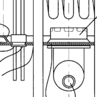
[0008] FIG. 1 is a block diagram of an imaging system for minimally invasive surgery.
【図1】図1は、低侵襲手術に対する画像診断システムのブロック図である。
US2021380693
In the occult stage, the cancer cannot be seen by imaging or bronchoscopy. In Stage 0, cancer cells are found in the lining of the airways.
潜伏ステージにおいて、画像診断や気管支鏡検査では癌を確認できない。ステージ0では、癌細胞が気道の内壁に見られる。
US10543179
In a specific embodiment of the invention, eye examinations include at least one of eye history, visual acuity, dilated ophthalmoscopy, Optical Coherence Tomography (OCT), evaluation of the fundus.
【0081】
本発明の特定の実施態様では、眼の検査としては、眼の病歴、視力、拡張検眼、光干渉断層画像診断(OCT)、眼底の検査のうちの少なくとも一つが挙げられる。
Such examinations are preferably performed by an ophthalmologist.
このような検査は好ましくは眼科医によって行う。
US10327790
[0124] As illustrated in FIG. 1, the arterial access device 2010 is transcervically introduced directly into the common carotid artery CCA of the patient.
図1に図示されているように、動脈アクセスデバイス2010は、患者の総頚動脈CCAへ経頚部的に直接導入される。
This may be done with a percutaneous puncture or a direct cut-down.
これは経皮的穿刺又は直接的な切開によって行うことができる。
In the case of a puncture, ultrasound imaging may be used to accurately make the initial arterial puncture.
穿刺の場合、最初の動脈穿刺を正確に行うために超音波画像診断を使用することができる。
US2021390764(JP)
[0003] With recent advancements in medical apparatuses including CT (computed tomography) apparatuses and MRI (magnetic resonance imaging) apparatuses, high-quality and high-resolution three-dimensional images have been increasingly used in diagnostic imaging.
【0002】
近年、CT(Computed Tomography)装置およびMRI(Magnetic Resonance Imaging)装置等の医療機器の進歩により、質の高い高解像度の3次元画像が画像診断に用いられるようになってきている。
US2022157453(JP)
[0047] In the operation room A, an operating place camera 10 , an operative field camera 11 , an endoscope system 12 , a video microscope system 13 , an ultrasound image diagnosis system 14 , a vital monitor 15 , a monitor 16 - 1 , a monitor 16 - 2 , an internet protocol (IP) switcher 17 , and a controller 18 are installed.
【0026】
手術室Aには、術場カメラ10、術野カメラ11、内視鏡システム12、ビデオ顕微鏡システム13、超音波画像診断システム14、バイタルモニタ15、モニタ16-1、モニタ16-2、IP(Internet Protocol)スイッチャ17、コントローラ18が設置されている。
US2021369947
[0004] Microparticles are employed by interventional radiologists for the selective occlusion of blood vessels in the treatment of, for example, hypervascular tumors such as leiomyoma uteri, and vascular anomalies such as vascular malformations.
【0004】
微粒子は、例えば、子宮平滑筋腫のような多血管性腫瘍、および血管奇形のような血管異常の治療における血管の選択的閉塞のために、インターベンショナルラジオロリス(画像診断医)によって使用される。
Such microparticles are injected into a hepatic artery of a patient.
このような微粒子は、患者の肝動脈に注入される。
US2010128049
The key problems in analyzing the radiological images are the sub-optimal conditions for review of digital radiology images
そのような放射線画像を分析する際の重要課題となるのは、デジタル放射線画像をレビューするには最適とは言えないそのような条件である。
that potentially results in diminished ability of radiologists and other clinicians to differentiate anatomy features in medical images,
これにより、画像診断医または他の臨床医の、医療画像における解剖学的特性を識別する能力が潜在的に低下する。
as well as increased eye fatigue and higher costs associated with diagnostic grade gray scale monitors.
また更に、診断に用いられるグレードのグレースケールモニタに高い費用が掛かるという問題や、目の疲れが増大するという問題もある。
US2021210206(JP)
[0045] The information about the past report is information about a report generated in the past with respect to the target image,
【0039】
過去のレポートの情報は、対象画像に対して過去に生成されたレポートの情報であり、
and information such as an image ID for identifying the target image, an image reading doctor ID for identifying an image diagnosis doctor who performed image reading, a lesion name, position information of the lesion, and findings are recorded.
対象画像を識別する画像ID、読影を行った画像診断医を識別するための読影医ID、病変名、病変の位置情報、及び所見等の情報が記録されている。
US9330455(JP)
With this apparatus, such diagnosis is possible in which a technician captures a test image and an image-based diagnostician at a distant location makes diagnosis while viewing the image of a lesion site and past case data similar to the lesion site displayed on the display device 1005 .
そこで、たとえば、技師が検査画像を撮像し、遠隔地にいる画像診断医が表示装置1005に表示された病変部の画像および病変部に類似する過去症例データを見ながら、診断を行うことができる。
US2020286234(JP)
[0049] The radiology report database 8
【0038】
読影レポートデータベース8には、
records radiology reports storing information such as, for example, an image ID identifying a medical image of a radiology object, a radiologist ID for identifying an image diagnostician interpreting the image, a lesion name, location information of the lesion, a finding, a confidence level of the finding, and the like.
例えば、読影対象の医用画像を識別する画像ID、読影を行った画像診断医を識別するための読影医ID、病変名、病変の位置情報、所見、所見の確信度等の情報が記録された読影レポートが登録される。