


05/15/2014 Tokyo Shimbun
To a husband's right lung and a wife's left lung It is a precision model making-transplant with 3D printer.
May 15, 2014 Morning paper
It makes with a
 3D printer, The model of the lung and chest which were used for prior examination of the living body lung transplantation.
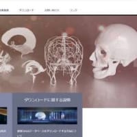
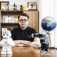
 The right is in the Professor Date Youji =14 day afternoon of the Kyoto University hospital, and Kyoto.
The Kyoto University hospital succeeded in the first living body lung transplantation in the world of transplanting a part of a husband's right lung as a left lung of the wife who is an incurable disease patient.
The hospital announced on the 14th.
It is 3D printer which played the large role in this operation.
Since it was the complicated operation of having made it rotating and transplanting the back and front of a lung, 3D printer was used in advance, the model with a lung or a precise blood vessel was made, and having examined the procedure led to a success.

The lung of the patient who received this living body lung transplantation is the 40th generation woman of the incurable disease "idiopathic interstitial pneumonia" which becomes hard gradually.
It examined excising the left lung which is following condition of disease, and transplanting the lower half of a husband's (his 40's) left lung at the beginning.
However, since the left-hand side where a human lung has the heart is small, it is judged that the transplant of the lower half is not enough as the lung capacity after an operation.
It decided to transplant a part of a husband's larger right lung as a left lung.
In a lung transplantation, it is required to sew up a bronchus, the pulmonary artery, and the pulmonary vein.
In order to turn over and transplant the lung of a right-and-left contrary this time, different how from usually to tie needed to be examined.
a hospital is a request about cooperation to the professors (medical design engineering) of the history of the Kunimoto Kei(and -- carrying out) of the department of the Nagoya City University graduate school art engineering research.
Professor Kunimoto reproduces the blood vessel of a husband's lung, and a wife's bronchus as a model faithfully with 3D printer based on the data of CAT.
The hospital repeated examination of the operation procedure by this model, and decided on different how from the former to tie.
The operation early in March advances as assumption.
By the time the wife who was bedridden mostly is good at housekeeping, she will recover, and she leaves hospital this month, and it is said that the husband is also doing the work return.
Kyoto University [ which performed an operation ] Professor Date Youji (a fathom -- carrying out) -- "-- the first operation has examined the procedure using the precise model -- a sake -- enforcement -- it has decided .
It is being said that the patient's inside of the body was as the model."
05/15/2014 東京新聞
夫の右肺、妻の左肺に 3Dプリンターで精密模型作り移植
2014年5月15日 朝刊
3Dプリンターで作り、生体肺移植の事前検討に利用した肺や胸部の模型。右は京都大病院の伊達洋至教授=14日午後、京都市で
夫の右肺の一部を、難病患者である妻の左肺として移植する世界で初めての生体肺移植に、京都大病院が成功した。病院が十四日発表した。この手術に大きな役割を果たしたのが3Dプリンター。肺の表裏を回転させて移植するという複雑な手術なので、事前に3Dプリンターを用いて肺や血管の精密な模型を作り、手順を検討したことが、成功につながった。
◆「患者臓器」複製し準備 京大肺移植世界初手術に効果
京都大病院が十四日発表した、左右逆の肺を移植する世界初の生体肺移植の成功には、3Dプリンター技術が大きな役割を果たした。ものづくりの可能性を広げている3Dプリンターは、外科での利用も進んでいるが、気管や血管が複雑に入り組んだ肺での活用は初めてという。 (森耕一)
3Dプリンターで作り、生体肺移植の事前検討に利用した肺や胸部の模型。右は京都大病院の伊達洋至教授=14日午後、京都市で
この生体肺移植を受けた患者は、肺が徐々に硬くなる難病「特発性間質性肺炎」の四十代女性。当初は、病状の進んでいる左肺を切除し、夫(四十代)の左肺の下半分を移植することを検討した。しかし、ヒトの肺は心臓のある左側は小さいため、その下半分の移植では手術後の肺活量が十分ではないと判断。より大きい夫の右肺の一部を、左肺として移植することにした。
肺移植では気管支、肺動脈、肺静脈を縫い合わせることが必要。今回は左右逆の肺を裏返して移植するため、通例と異なるつなぎ方を検討する必要があった。
病院は、名古屋市立大大学院芸術工学研究科の國本桂史(かつし)教授(医療デザイン工学)らに協力を依頼。國本教授はCTスキャンのデータをもとに夫の肺の血管や妻の気管支を3Dプリンターで忠実に模型として再現。病院は、この模型で手術手順の検討を重ね、従来とは異なるつなぎ方を決めた。
三月上旬に実施した手術は想定通りに進行。ほぼ寝たきりだった妻は家事ができるまでに回復して今月退院し、夫も仕事復帰しているという。執刀した京大の伊達洋至(ひろし)教授は「初めての手術で、緻密な模型を使って手順を検討できたため、実施を決断できた。患者の体内は模型通りだった」と話している。


※コメント投稿者のブログIDはブログ作成者のみに通知されます