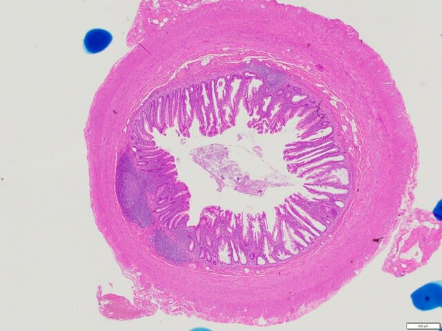
病理と臨床の2021年2月号「今月の話題」(自己免疫性胃炎の初期像に迫る)から。早期の自己免疫性胃炎の組織学的特徴?(下記文献と教科書を参考にしています)
1. びまん性または巣状に密なリンパ球・形質細胞浸潤が粘膜深層優位にみられる。
2. リンパ球はCD4(+)が主体で、好酸球も混じる。
3. リンパ球は胃底腺破壊性に浸潤し、アポトーシス体が観察される。
4. 偽幽門腺・幽門腺化生が生じるが、腸上皮化生と膵上皮化生は早期ではまれである。
5. 壁細胞突出 parietal cell protrusion (PCP) がみられる。プロトンポンプ阻害薬でいつもみるような刺々しさはなく弱々しい印象を受ける。
6. ECL細胞の線状過形成 linear hyperplasiaが認められるが、nodular hyperplasia (ECM) はまれである。クロモグラニンA染色が推奨される(この場合、シナプトフィジン染色はおすすめできません)。
参照文献
・Torbenson, M., et al.: Autoimmune gastritis: distinct histological and immunohistochemical findings before complete loss of oxyntic glands Mod Pathol 2002, 15: 102-109
・Greenson, J.K., Lawers, G.Y., Montgomery, E.A. (eds.): Diagnostic pathology; Gastrointestinal. 3rd ed. Elsevier, Amsterdam, 2019
*お詫び:「今月の話題」の図1説明文でparietal cell protrusionの略語がPCAとなっていますが、PCPの間違いです。今度、訂正文が入る予定です。
Pathology and Clinical Medicine (February 2021): "Topic of the Month"
Histological features of early-phase autoimmune gastritis? (Based on the following literature and textbook)
1. Diffuse or focal dense lymphocytic and plasma cell infiltrates, predominate in the deeper lamina propria mucosae.
2. Lymphocytes are predominantly CD4(+) with eosinophils.
3. Lymphocytes infiltrate the oxyntic glands destructively with apoptotic bodies.
4. Pseudo-pyloric and pyloric metaplasia occur, but intestinal and pancreatic metaplasia are rarely seen in the early stage.
5. Parietal cell protrusion (PCP) is seen, but looks weaker. PCP is not as sharp as that usually seen with proton pump inhibitors.
6. Linear hyperplasia of ECL-cells is seen, while the nodular hyperplasia (so-called ECM) is rare. Chromogranin A staining is recommended. Synaptophysin staining is not recommended in this situation.
・Torbenson, M., et al.: Autoimmune gastritis: distinct histological and immunohistochemical findings before complete loss of oxyntic glands Mod Pathol 2002, 15: 102-109
・Greenson, J.K., Lawers, G.Y., Montgomery, E.A. (eds.): Diagnostic pathology; Gastrointestinal. 3rd ed. Elsevier, Amsterdam, 2019
静岡県の先生がリモート講演で「弱々しいPCP」を使っていただいたので、お近くの名所「家山桜」を見に行ってきました。
A famous endoscopist from Shizuoka Prefecture used "Weaker PCP" in a remote lecture, so I went to see "Ieyama Cherry Blossom" along the Oigawa Railway.

2021.3.20.家山川橋梁にて
C11 190 crossing Ieyama River, Shizuoka, Japan.