・ 科学雑誌編集者・出版社への申立て
申立てPart.1, 申立てPart.2, 申立てPart.3, 申立てPart.4
・ 申立てに対する、科学雑誌編集者・出版社からの返信メール
返信Part.1、返信Part.2、返信Part.3
Life Sciences誌への申立て
件名
Allegations of fabrication and falsification (Life Sciences 2008;82:884-91).
宛先
frankp@email.arizona.edu
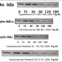
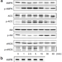
Please investigate fraudulent data of the article “PPARalpha activators upregulate eNOS activity and inhibit cytokine-induced NF-kappaB activation through AMP-activated protein kinase activation.” Okayasu T, Tomizawa A, Suzuki K, Manaka K, Hattori Y., published in Life Sciences (2008;82,884-91). You can find fabricated (falsified) Fig. 1(A),(B) and Fig. 5(A),(B),(C) : namely,
(1)
The Phospho-IkB-alpha image in Fig. 4(A) of Ref.2 is similar to
the Phospho-IkB-alpha image in Fig. 5(A) of Ref.3
and the Phospho-IkBa image in Fig. 3(A) of Ref.4
and the Phospho IkB-alpha image in Fig. 6(A) of Ref.5.
(2)
The IkB-alpha image in Fig. 4(B) of Ref.2 is similar to
the IKB-alpha image in Fig. 5(B) of Ref.3
and the IkBa image in Fig. 3(B) of Ref.4
and the leftside 4 bands of the ONOO-IkB-alpha image in Fig. 6(A) of Ref.5.
(3)
The GST-Ikb-alpha image of top panel in Fig. 4(C) of Ref.1 is similar to both GST-Ikb-alpha image of top panel in Fig.4(C) of Ref.2 and IkB-alpha image of top panel in Fig. 5(C) of Ref.3
(4)
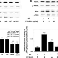
The IKKalpha/beta image in Fig. 4(C) of Ref.2 is similar to both the ACC image in Fig. 1(A) of Ref.3 and the IKKalpha/beta image in Fig. 5(C) of Ref.3.
(5)
The GST-Ikb-alpha image of bottom panel in Fig. 4(C) of Ref.2 is similar to GST-Ikb-alpha image of bottom panel in Fig.4(C) of Ref.3. Moreover, these two images are similar to both the left-right reversal of the Akt image in Fig. 1(A) of Ref.1 and the upside-down + left-right reversal of the GST-Ikb-alpha image of bottom panel in Fig.4(C) of Ref.1
(6)
The GST-IkB-alpha image, bottom panel in Fig. 4(C) of Ref.2 is similar to the Akt image in Fig. 1(A) of Ref.3.
(7)
The p-AKT image in Fig. 1(A) of Ref.3 is similar to the p-ACC image in Fig. 1(B) of Ref.3
(8)
The IkB-alpha image in Fig. 5(A) of Ref.3 is similar to both the IkB-a image in Fig. 3(A) of Ref.4 and the IkB-alpha image in Fig. 6(A) of Ref.5.
(9)
The AMPK and pAMPK images in Fig. 1(A) of Ref.1 are similar to the AMPK and pAMPK images in Fig. 1(A) of Ref.3.
(10)
The p-AKT image in Fig. 1(A) of Ref.1 is similar to the p-Akt image in Fig. 1(A) of Ref.3.
(11)
The eNOS image in Fig. 1(A) of Ref.1 is similar to the eNOS image in Fig. 1(A) of Ref.3.
References
1,
Metabolism. 2010 Jun 25. [Epub ahead of print]
Fenofibrate suppresses microvascular inflammation and apoptosis through adenosine monophosphate-activated protein kinase activation.
Tomizawa A, Hattori Y, Inoue T, Hattori S, Kasai K.
2,
Cardiovasc Res. 2009 Jan 1;81(1):133-9.
3,
Life Sci. 2008 Apr 9;82(15-16):884-91.
4,
Hypertension. 2006 Jun;47(6):1183-8.
5,
Cardiovasc Res. 2004 Jul 1;63(1):31-40.
Hypertension誌への申立て
件名
Allegations of fabrication and falsification (Hypertension 2006 Jun;47(6):1183-8.)
宛先
hypertension@physiology.umsmed.edu ; jehall@physiology.umsmed.edu
CC
hypertension@umc.edu
Dear Dr.John E. Hall, Editor-in-Chief of Hypertension,
Please investigate fraudulent data of the article “Metformin inhibits Cytokine-Induced NF-kB Activation via AMPK Activation in Vascular Endothelial Cells” Hattori Y, Suzuki K, Hattori S, Kasai K., published in Hypertension (2006 Jun;47(6):1183-8.). You can find fabricated (falsified) Fig. 3(A),(B),(C) and Fig. 4(B) : namely,
(1)
The Phospho-IkB-alpha image in Fig. 4(A) of Ref.1 is similar to
the Phospho-IkB-alpha image in Fig. 5(A) of Ref.2
and the Phospho-IkBa image in Fig. 3(A) of Ref.4
and the Phospho IkB-alpha image in Fig. 6(A) of Ref.6.
(2)
The IkB-alpha image in Fig. 4(B) of Ref.1 is similar to
the IKB-alpha image in Fig. 5(B) of Ref.2
and the IkBa image in Fig. 3(B) of Ref.4
and the leftside 4 bands of the ONOO-IkB-alpha image in Fig. 6(A) of Ref.6.
(3)
The IkB-alpha image in Fig. 5(A) of Ref.2 is similar to both the IkB-a image in Fig. 3(A) of Ref.4 and the IkB-alpha image in Fig. 6(A) of Ref.6.
(4)
The right-side 3 bands (second, third and fourth bands from left) of IkB-alpha image, top-panel in Fig. 3(C) of Ref.4 is similar to the middle 3 bands (second, third and fourth bands from left) of IkB-alpha image, top-panel in Fig. 6(B) of Ref.6.
(5)
The IKKa/b image in Fig. 3(C) of Ref.4 is similar to the left-side 4 bands (first to fourth bands from left) of the IKKalpha/beta image, fifth panel from bottom in Fig. 6(B) of Ref.6.
Moreover, the left-side 3 bands of these two images are similar to the IKKalpha/beta image, second panel from bottom in Fig. 6(B) of Ref.6.
(6)
The IkB-a image, bottom panel in Fig. 3(C) of Ref.4 is similar to the left-side 4 bands (first to fourth bands from left) of IkB-alpha image, fourth panel from bottom in Fig. 6(B) of Ref.6.
Moreover, the left-side 3 bands of these image are similar to the IkB-alpha image, bottom panel in Fig. 6(B) of Ref.6.
(7)
The eNOS image in Fig. 4(A) of Ref.3 is similar to the right-side 6 bands (for BH4 only, AngII, AngII/BH4) of eNOS Protein image in Fig. 5(a) of Ref.5.
Moreover, these two images are similar to the 6 bands (third to eighth bands from left) of the MCP-1 image in Fig. 4(B) of Ref.4.
References
1
Cardiovasc Res. 2009 Jan 1;81(1):133-9.
2,
Life Sci. 2008 Apr 9;82(15-16):884-91.
3,
Eur J Pharmacol. 2007 Jan 19;555(1):48-53.
4,
Hypertension. 2006 Jun;47(6):1183-8.
5
J Hypertens. 2005 Jul;23(7):1375-82.
6
Cardiovasc Res. 2004 Jul 1;63(1):31-40.
Cardiovasc Research誌への申立て
件名
Allegations of fabrication and falsification (Cardiovasc Research 2004 Jul 1;63(1):31-40)
宛先
CVR@physiologie.med.uni-giessen.de
Dear Dr. Hans Michael Piper,
Please investigate fraudulent data of the article “NO suppresses while peroxynitrite sustains NF-kB: A paradigm to rationalize cytoprotective and cytotoxic actions attributed to NO” Hattori Y, Kasai K, Gross SS, published in Cardiovascular Research. (2004 Jul 1;63(1):31-40.). You can find fabricated (falsified) Fig. 6(A),(B) : namely,
(1)
The Phospho-IkB-alpha image in Fig. 4(A) of Ref.1 is similar to
the Phospho-IkB-alpha image in Fig. 5(A) of Ref.2
and the Phospho-IkBa image in Fig. 3(A) of Ref.3
and the Phospho IkB-alpha image in Fig. 6(A) of Ref.4.
(2)
The IkB-alpha image in Fig. 4(B) of Ref.1 is similar to
the IKB-alpha image in Fig. 5(B) of Ref.2
and the IkBa image in Fig. 3(B) of Ref.3
and the leftside 4 bands of the ONOO-IkB-alpha image in Fig. 6(A) of Ref.4.
(3)
The IkB-alpha image in Fig. 5(A) of Ref.2 is similar to both the IkB-a image in Fig. 3(A) of Ref.3 and the IkB-alpha image in Fig. 6(A) of Ref.4.
(4)
The right-side 3 bands (second, third and fourth bands from left) of IkB-alpha image, top-panel in Fig. 3(C) of Ref.3 is similar to the middle 3 bands (second, third and fourth bands from left) of IkB-alpha image, top-panel in Fig. 6(B) of Ref.4.
(5)
The second band from left of IkB-alpha image, top-panel in Fig. 6(B) of Ref.4 is similar to the second band from left of IkB-alpha image, third panel from bottom in Fig. 6(B) of Ref.4.
(6)
The IKKa/b image in Fig. 3(C) of Ref.3 is similar to the left-side 4 bands (first to fourth bands from left) of the IKKalpha/beta image, fifth panel from bottom in Fig. 6(B) of Ref.4.
Moreover, the left-side 3 bands of these two images are similar to the IKKalpha/beta image, second panel from bottom in Fig. 6(B) of Ref.4.
(7)
The IkB-a image, bottom panel in Fig. 3(C) of Ref.3 is similar to the left-side 4 bands (first to fourth bands from left) of IkB-alpha image, fourth panel from bottom in Fig. 6(B) of Ref.4.
Moreover, the left-side 3 bands of these image are similar to the IkB-alpha image, bottom panel in Fig. 6(B) of Ref.4.
(8)
The left-side 3 bands (first, second and third bands from left) of NOR3 IkB-alpha image in Fig. 6(A) of Ref.4 is similar to the right-side 3 bands (third, fourth and fifth bands from left) of IKKalpha/beta image, fifth panel from bottom in Fig. 6(B) of Ref.4.
References
1
Cardiovasc Res. 2009 Jan 1;81(1):133-9.
2,
Life Sci. 2008 Apr 9;82(15-16):884-91.
3,
Hypertension. 2006 Jun;47(6):1183-8.
4,
Cardiovasc Res. 2004 Jul 1;63(1):31-40.
JAT誌への申立て
件名
Allegations of fabrication and falsification (JAT Vol. 17 (2010) , No. 5 503-509.).
宛先
shinichi@nms.ac.jp
CC
jas@kk-kyowa.co.jp
Please investigate fraudulent data of the article “Anti-inflammatory role of cilostazol in vascular smooth muscle cells in vitro and in vivo.” Aoki C, Hattori Y, Tomizawa A, Jojima T, Kasai K., published in Journal of Atherosclerosis and Thrombosis. (Vol. 17 (2010) , No. 5 503-509.). You can find fabricated (falsified) Fig. 2(A),(B) and 3(A) : namely,
(1)
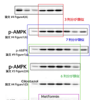
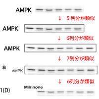
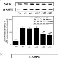
The first, second and third bands from right of IkB-alpha image, top-panel in Fig. 4(A) of Ref.2 is similar to the second, third and fourth bands from left of pAMPK image in Fig. 1(A) of Ref.2.
Moreover, the first and second bands from left of IkB-alpha image, top-panel in Fig. 4(A) of Ref.2 is similar to both the fifth and sixth bands from left of pAMPK image in Fig. 1(A) of Ref.2
and the fifth and sixth bands from left of pAMPK image in Fig. 1(a) of Ref.4.
Furthermore, the pAMPK image (all 7 bands) in Fig. 1(A) of Ref.2 is similar to the pAMPK image (all 7 bands) in Fig. 1(a) of Ref.4
,and first to sixth bands from left of the above pAMPK image of Ref.2,9 are similar to the pAMPK image for cilostazol in Fig. 1(D) of Ref.2
and the pAMPK image in Fig. 2(A) of Ref.1
and the pAMPK image for Metformin in Fig. 3(b) of Ref.3.
In addition, the rightside 5 bands (2.5 min – 60 min) of the pAMPK imgae in Fig. 2(A) of Ref.1 are similar to upside-down and left-right reversal of the pAMPK image in Fig. 3(A) of Ref.1.
(2)
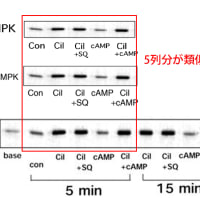
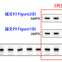
The AMPK image in Fig. 1(A) of Ref.2 is similar to the APMK image in Fig. 1(a) of Ref.4.
Moreover, the leftside 6 bands of these two APMK image are similar to the AMPK image in Fig. 2(A) of Ref.1.
Furthermore, rightside 5 bands of this AMPK image in Fig. 2(A) of Ref.1 are similar to the pAMPK image for Milrinone in Fig. 1(D) of Ref.2
(3)
The rightside 5 bands of the AMPK image in Fig. 1(B) of Ref.2 is similar to both the AMPK image in Fig. 2(B) of Ref.1 and the AMPK image in Fig. 1(b) of Ref.4.
Likewise, 5 bands (second to sixth bands from left) of pAMPK image in Fig. 1(B) of Ref.2 are similar to the pAMPK image in Fig. 2(B) of Ref.1 and the pAMPK image in Fig. 1(b) of Ref.4.
(4)
The ACC and pACC images in Fig. 1(A) of Ref.2 are similar to the ACC and pACC images in Fig. 1(a) of Ref.4, respectively.
Moreover, the leftside 6 bands of these two images are similar to the ACC and p-ACC images in Fig. 2(A) of Ref.1.
References
1,
J Atheroscler Thromb. 2010;17(5):503-9.
2,
Cardiovasc Res. 2009 Jan 1;81(1):133-9.
3,
Hypertens Res. 2009 Sep;32(9):765-9.
4.
Am J Hypertens. 2008 Apr;21(4):451-7.
Hypertension Res誌への申立て
件名
Allegations of fabrication and falsification (Hypertension Res 2009 ;32:765-9)
宛先
horiuchi@m.ehime-u.ac.jp
CC
kkario@jichi.ac.jp ; mmogi@m.ehime-u.ac.jp
Please investigate fraudulent data of the article “Telmisartan inhibits cytokine-induced nuclear factor-kappaB activation independently of the peroxisome proliferator-activated receptor-gamma. Nakano A, Hattori Y, Aoki C, Jojima T, Kasai K., published in Hypertens Research. (2009 Sep;32(9):765-9.). You can find fabricated (falsified) Fig. 3(b) : namely,
(1)
The right-side 3 bands (first, second and third bands from right) of IkB-alpha image, top-panel in Fig. 4(A) of Ref.2 are similar to the second, third and fourth bands from left of pAMPK image in Fig. 1(A) of Ref.2.
Moreover, the first and second bands from left of IkB-alpha image, top-panel in Fig. 4(A) of Ref.2 is similar to both the fifth and sixth bands from left of pAMPK image in Fig. 1(A) of Ref.2
and the fifth and sixth bands from left of pAMPK image in Fig. 1(a) of Ref.4.
Furthermore, the pAMPK image (all 7 bands) in Fig. 1(A) of Ref.2 is similar to the pAMPK image (all 7 bands) in Fig. 1(a) of Ref.4
,and first to sixth bands from left of the above pAMPK image of Ref.2,9 are similar to the pAMPK image for cilostazol in Fig. 1(D) of Ref.2
and the pAMPK image in Fig. 2(A) of Ref.1
and the pAMPK image for Metformin in Fig. 3(b) of Ref.3.
In addition, the rightside 5 bands (2.5 min – 60 min) of the pAMPK imgae in Fig. 2(A) of Ref.1 are similar to upside-down and left-right reversal of the pAMPK image in Fig. 3(A) of Ref.1.
References
1,
J Atheroscler Thromb. 2010;17(5):503-9.
2,
Cardiovasc Res. 2009 Jan 1;81(1):133-9.
3,
Hypertens Res. 2009 Sep;32(9):765-9.
4,
Am J Hypertens. 2008 Apr;21(4):451-7.
FEBS Letters誌への申立て
件名
Allegations of fabrication and falsification (FEBS Lett. 2008;582:1719-24)
宛先
febs.letters@bzh.uni-heidelberg.de
CC
felix.wieland@bzh.uni-heidelberg.de ;
Please investigate fraudulent data of the article “High molecular weight adiponectin activates AMPK and suppresses cytokine-induced NF-κB activation in vascular endothelial cells” Hattori Y, Nakano Y, Hattori S, Tomizawa A, Inukai K, Kasai K., published in FEBS Letters. ( 2008 May 28;582(12):1719-24.). You can find fabricated (falsified) Fig. 2(A) : namely,
指摘項目No.19
The left-side 4 bands (first to fourth bands from left) of the AMPK image in Fig. 1(C) of Ref.1 is similar to the left-side 4 bands (first to fourth bands from left) of the AMPK image in Fig. 2(A) of Ref.2.
指摘項目No.20
The left-side 3 bands (first to third bands from left) of pAMPK image in Fig. 1(C) of Ref.1 is similar to the left-side 3 bands (firtst to third bands from left) of pAPMK image in Fig. 2(A) of Ref.2.
References
1,
Cardiovasc Res. 2009 Jan 1;81(1):133-9.
2,
FEBS Lett. 2008 May 28;582(12):1719-24.
Sincerely yours,
Juuichi Jigen
E-mail: 11jigen@mail.goo.ne.jp
Am J Hypertens誌への申立て
件名
Allegations of fabrication and falsification (Am J Hypertens. 2008;21:451-7)
宛先
yvonne.raiford@einstein.yu.edu
Please investigate fraudulent data of the article “Cilostazol activates AMP-activated protein kinase and restores endothelial function in diabetes.” By Suzuki K, Uchida K, Nakanishi N, Hattori Y., published in American Journal of Hypertension (2008 Apr;21(4):451-7.). You can find fabricated (falsified) Fig. 1(a),(b) : namely,
(1)
The first, second and third bands from right of IkB-alpha image, top-panel in Fig. 4(A) of Ref.2 is similar to the second, third and fourth bands from left of pAMPK image in Fig. 1(A) of Ref.2.
Moreover, the first and second bands from left of IkB-alpha image, top-panel in Fig. 4(A) of Ref.2 is similar to both the fifth and sixth bands from left of pAMPK image in Fig. 1(A) of Ref.2
and the fifth and sixth bands from left of pAMPK image in Fig. 1(a) of Ref.4.
Furthermore, the pAMPK image (all 7 bands) in Fig. 1(A) of Ref.2 is similar to the pAMPK image (all 7 bands) in Fig. 1(a) of Ref.4
,and first to sixth bands from left of the above pAMPK image of Ref.2,9 are similar to the pAMPK image for cilostazol in Fig. 1(D) of Ref.2
and the pAMPK image in Fig. 2(A) of Ref.1
and the pAMPK image for Metformin in Fig. 3(b) of Ref.3.
In addition, the rightside 5 bands (2.5 min – 60 min) of the pAMPK imgae in Fig. 2(A) of Ref.1 are similar to upside-down and left-right reversal of the pAMPK image in Fig. 3(A) of Ref.1.
(2)
The AMPK image in Fig. 1(A) of Ref.2 is similar to the APMK image in Fig. 1(a) of Ref.4.
Moreover, the leftside 6 bands of these two APMK image are similar to the AMPK image in Fig. 2(A) of Ref.1.
Furthermore, rightside 5 bands of this AMPK image in Fig. 2(A) of Ref.1 are similar to the pAMPK image for Milrinone in Fig. 1(D) of Ref.2
(3)
The rightside 5 bands of the AMPK image in Fig. 1(B) of Ref.2 is similar to both the AMPK image in Fig. 2(B) of Ref.1 and the AMPK image in Fig. 1(b) of Ref.4.
Likewise, 5 bands (second to sixth bands from left) of pAMPK image in Fig. 1(B) of Ref.2 are similar to the pAMPK image in Fig. 2(B) of Ref.1 and the pAMPK image in Fig. 1(b) of Ref.4.
(4)
The ACC and pACC images in Fig. 1(A) of Ref.2 are similar to the ACC and pACC images in Fig. 1(a) of Ref.4, respectively.
Moreover, the leftside 6 bands of these two images are similar to the ACC and p-ACC images in Fig. 2(A) of Ref.1.
References
1,
J Atheroscler Thromb. 2010;17(5):503-9.
2,
Cardiovasc Res. 2009 Jan 1;81(1):133-9.
3,
Hypertens Res. 2009 Sep;32(9):765-9.
4.
Am J Hypertens. 2008 Apr;21(4):451-7.
申立てPart.1, 申立てPart.2, 申立てPart.3, 申立てPart.4
・ 申立てに対する、科学雑誌編集者・出版社からの返信メール
返信Part.1、返信Part.2、返信Part.3
Life Sciences誌への申立て
件名
Allegations of fabrication and falsification (Life Sciences 2008;82:884-91).
宛先
frankp@email.arizona.edu
Please investigate fraudulent data of the article “PPARalpha activators upregulate eNOS activity and inhibit cytokine-induced NF-kappaB activation through AMP-activated protein kinase activation.” Okayasu T, Tomizawa A, Suzuki K, Manaka K, Hattori Y., published in Life Sciences (2008;82,884-91). You can find fabricated (falsified) Fig. 1(A),(B) and Fig. 5(A),(B),(C) : namely,
(1)
The Phospho-IkB-alpha image in Fig. 4(A) of Ref.2 is similar to
the Phospho-IkB-alpha image in Fig. 5(A) of Ref.3
and the Phospho-IkBa image in Fig. 3(A) of Ref.4
and the Phospho IkB-alpha image in Fig. 6(A) of Ref.5.
(2)
The IkB-alpha image in Fig. 4(B) of Ref.2 is similar to
the IKB-alpha image in Fig. 5(B) of Ref.3
and the IkBa image in Fig. 3(B) of Ref.4
and the leftside 4 bands of the ONOO-IkB-alpha image in Fig. 6(A) of Ref.5.
(3)
The GST-Ikb-alpha image of top panel in Fig. 4(C) of Ref.1 is similar to both GST-Ikb-alpha image of top panel in Fig.4(C) of Ref.2 and IkB-alpha image of top panel in Fig. 5(C) of Ref.3
(4)
The IKKalpha/beta image in Fig. 4(C) of Ref.2 is similar to both the ACC image in Fig. 1(A) of Ref.3 and the IKKalpha/beta image in Fig. 5(C) of Ref.3.
(5)
The GST-Ikb-alpha image of bottom panel in Fig. 4(C) of Ref.2 is similar to GST-Ikb-alpha image of bottom panel in Fig.4(C) of Ref.3. Moreover, these two images are similar to both the left-right reversal of the Akt image in Fig. 1(A) of Ref.1 and the upside-down + left-right reversal of the GST-Ikb-alpha image of bottom panel in Fig.4(C) of Ref.1
(6)
The GST-IkB-alpha image, bottom panel in Fig. 4(C) of Ref.2 is similar to the Akt image in Fig. 1(A) of Ref.3.
(7)
The p-AKT image in Fig. 1(A) of Ref.3 is similar to the p-ACC image in Fig. 1(B) of Ref.3
(8)
The IkB-alpha image in Fig. 5(A) of Ref.3 is similar to both the IkB-a image in Fig. 3(A) of Ref.4 and the IkB-alpha image in Fig. 6(A) of Ref.5.
(9)
The AMPK and pAMPK images in Fig. 1(A) of Ref.1 are similar to the AMPK and pAMPK images in Fig. 1(A) of Ref.3.
(10)
The p-AKT image in Fig. 1(A) of Ref.1 is similar to the p-Akt image in Fig. 1(A) of Ref.3.
(11)
The eNOS image in Fig. 1(A) of Ref.1 is similar to the eNOS image in Fig. 1(A) of Ref.3.
References
1,
Metabolism. 2010 Jun 25. [Epub ahead of print]
Fenofibrate suppresses microvascular inflammation and apoptosis through adenosine monophosphate-activated protein kinase activation.
Tomizawa A, Hattori Y, Inoue T, Hattori S, Kasai K.
2,
Cardiovasc Res. 2009 Jan 1;81(1):133-9.
3,
Life Sci. 2008 Apr 9;82(15-16):884-91.
4,
Hypertension. 2006 Jun;47(6):1183-8.
5,
Cardiovasc Res. 2004 Jul 1;63(1):31-40.
Hypertension誌への申立て
件名
Allegations of fabrication and falsification (Hypertension 2006 Jun;47(6):1183-8.)
宛先
hypertension@physiology.umsmed.edu ; jehall@physiology.umsmed.edu
CC
hypertension@umc.edu
Dear Dr.John E. Hall, Editor-in-Chief of Hypertension,
Please investigate fraudulent data of the article “Metformin inhibits Cytokine-Induced NF-kB Activation via AMPK Activation in Vascular Endothelial Cells” Hattori Y, Suzuki K, Hattori S, Kasai K., published in Hypertension (2006 Jun;47(6):1183-8.). You can find fabricated (falsified) Fig. 3(A),(B),(C) and Fig. 4(B) : namely,
(1)
The Phospho-IkB-alpha image in Fig. 4(A) of Ref.1 is similar to
the Phospho-IkB-alpha image in Fig. 5(A) of Ref.2
and the Phospho-IkBa image in Fig. 3(A) of Ref.4
and the Phospho IkB-alpha image in Fig. 6(A) of Ref.6.
(2)
The IkB-alpha image in Fig. 4(B) of Ref.1 is similar to
the IKB-alpha image in Fig. 5(B) of Ref.2
and the IkBa image in Fig. 3(B) of Ref.4
and the leftside 4 bands of the ONOO-IkB-alpha image in Fig. 6(A) of Ref.6.
(3)
The IkB-alpha image in Fig. 5(A) of Ref.2 is similar to both the IkB-a image in Fig. 3(A) of Ref.4 and the IkB-alpha image in Fig. 6(A) of Ref.6.
(4)
The right-side 3 bands (second, third and fourth bands from left) of IkB-alpha image, top-panel in Fig. 3(C) of Ref.4 is similar to the middle 3 bands (second, third and fourth bands from left) of IkB-alpha image, top-panel in Fig. 6(B) of Ref.6.
(5)
The IKKa/b image in Fig. 3(C) of Ref.4 is similar to the left-side 4 bands (first to fourth bands from left) of the IKKalpha/beta image, fifth panel from bottom in Fig. 6(B) of Ref.6.
Moreover, the left-side 3 bands of these two images are similar to the IKKalpha/beta image, second panel from bottom in Fig. 6(B) of Ref.6.
(6)
The IkB-a image, bottom panel in Fig. 3(C) of Ref.4 is similar to the left-side 4 bands (first to fourth bands from left) of IkB-alpha image, fourth panel from bottom in Fig. 6(B) of Ref.6.
Moreover, the left-side 3 bands of these image are similar to the IkB-alpha image, bottom panel in Fig. 6(B) of Ref.6.
(7)
The eNOS image in Fig. 4(A) of Ref.3 is similar to the right-side 6 bands (for BH4 only, AngII, AngII/BH4) of eNOS Protein image in Fig. 5(a) of Ref.5.
Moreover, these two images are similar to the 6 bands (third to eighth bands from left) of the MCP-1 image in Fig. 4(B) of Ref.4.
References
1
Cardiovasc Res. 2009 Jan 1;81(1):133-9.
2,
Life Sci. 2008 Apr 9;82(15-16):884-91.
3,
Eur J Pharmacol. 2007 Jan 19;555(1):48-53.
4,
Hypertension. 2006 Jun;47(6):1183-8.
5
J Hypertens. 2005 Jul;23(7):1375-82.
6
Cardiovasc Res. 2004 Jul 1;63(1):31-40.
Cardiovasc Research誌への申立て
件名
Allegations of fabrication and falsification (Cardiovasc Research 2004 Jul 1;63(1):31-40)
宛先
CVR@physiologie.med.uni-giessen.de
Dear Dr. Hans Michael Piper,
Please investigate fraudulent data of the article “NO suppresses while peroxynitrite sustains NF-kB: A paradigm to rationalize cytoprotective and cytotoxic actions attributed to NO” Hattori Y, Kasai K, Gross SS, published in Cardiovascular Research. (2004 Jul 1;63(1):31-40.). You can find fabricated (falsified) Fig. 6(A),(B) : namely,
(1)
The Phospho-IkB-alpha image in Fig. 4(A) of Ref.1 is similar to
the Phospho-IkB-alpha image in Fig. 5(A) of Ref.2
and the Phospho-IkBa image in Fig. 3(A) of Ref.3
and the Phospho IkB-alpha image in Fig. 6(A) of Ref.4.
(2)
The IkB-alpha image in Fig. 4(B) of Ref.1 is similar to
the IKB-alpha image in Fig. 5(B) of Ref.2
and the IkBa image in Fig. 3(B) of Ref.3
and the leftside 4 bands of the ONOO-IkB-alpha image in Fig. 6(A) of Ref.4.
(3)
The IkB-alpha image in Fig. 5(A) of Ref.2 is similar to both the IkB-a image in Fig. 3(A) of Ref.3 and the IkB-alpha image in Fig. 6(A) of Ref.4.
(4)
The right-side 3 bands (second, third and fourth bands from left) of IkB-alpha image, top-panel in Fig. 3(C) of Ref.3 is similar to the middle 3 bands (second, third and fourth bands from left) of IkB-alpha image, top-panel in Fig. 6(B) of Ref.4.
(5)
The second band from left of IkB-alpha image, top-panel in Fig. 6(B) of Ref.4 is similar to the second band from left of IkB-alpha image, third panel from bottom in Fig. 6(B) of Ref.4.
(6)
The IKKa/b image in Fig. 3(C) of Ref.3 is similar to the left-side 4 bands (first to fourth bands from left) of the IKKalpha/beta image, fifth panel from bottom in Fig. 6(B) of Ref.4.
Moreover, the left-side 3 bands of these two images are similar to the IKKalpha/beta image, second panel from bottom in Fig. 6(B) of Ref.4.
(7)
The IkB-a image, bottom panel in Fig. 3(C) of Ref.3 is similar to the left-side 4 bands (first to fourth bands from left) of IkB-alpha image, fourth panel from bottom in Fig. 6(B) of Ref.4.
Moreover, the left-side 3 bands of these image are similar to the IkB-alpha image, bottom panel in Fig. 6(B) of Ref.4.
(8)
The left-side 3 bands (first, second and third bands from left) of NOR3 IkB-alpha image in Fig. 6(A) of Ref.4 is similar to the right-side 3 bands (third, fourth and fifth bands from left) of IKKalpha/beta image, fifth panel from bottom in Fig. 6(B) of Ref.4.
References
1
Cardiovasc Res. 2009 Jan 1;81(1):133-9.
2,
Life Sci. 2008 Apr 9;82(15-16):884-91.
3,
Hypertension. 2006 Jun;47(6):1183-8.
4,
Cardiovasc Res. 2004 Jul 1;63(1):31-40.
JAT誌への申立て
件名
Allegations of fabrication and falsification (JAT Vol. 17 (2010) , No. 5 503-509.).
宛先
shinichi@nms.ac.jp
CC
jas@kk-kyowa.co.jp
Please investigate fraudulent data of the article “Anti-inflammatory role of cilostazol in vascular smooth muscle cells in vitro and in vivo.” Aoki C, Hattori Y, Tomizawa A, Jojima T, Kasai K., published in Journal of Atherosclerosis and Thrombosis. (Vol. 17 (2010) , No. 5 503-509.). You can find fabricated (falsified) Fig. 2(A),(B) and 3(A) : namely,
(1)
The first, second and third bands from right of IkB-alpha image, top-panel in Fig. 4(A) of Ref.2 is similar to the second, third and fourth bands from left of pAMPK image in Fig. 1(A) of Ref.2.
Moreover, the first and second bands from left of IkB-alpha image, top-panel in Fig. 4(A) of Ref.2 is similar to both the fifth and sixth bands from left of pAMPK image in Fig. 1(A) of Ref.2
and the fifth and sixth bands from left of pAMPK image in Fig. 1(a) of Ref.4.
Furthermore, the pAMPK image (all 7 bands) in Fig. 1(A) of Ref.2 is similar to the pAMPK image (all 7 bands) in Fig. 1(a) of Ref.4
,and first to sixth bands from left of the above pAMPK image of Ref.2,9 are similar to the pAMPK image for cilostazol in Fig. 1(D) of Ref.2
and the pAMPK image in Fig. 2(A) of Ref.1
and the pAMPK image for Metformin in Fig. 3(b) of Ref.3.
In addition, the rightside 5 bands (2.5 min – 60 min) of the pAMPK imgae in Fig. 2(A) of Ref.1 are similar to upside-down and left-right reversal of the pAMPK image in Fig. 3(A) of Ref.1.
(2)
The AMPK image in Fig. 1(A) of Ref.2 is similar to the APMK image in Fig. 1(a) of Ref.4.
Moreover, the leftside 6 bands of these two APMK image are similar to the AMPK image in Fig. 2(A) of Ref.1.
Furthermore, rightside 5 bands of this AMPK image in Fig. 2(A) of Ref.1 are similar to the pAMPK image for Milrinone in Fig. 1(D) of Ref.2
(3)
The rightside 5 bands of the AMPK image in Fig. 1(B) of Ref.2 is similar to both the AMPK image in Fig. 2(B) of Ref.1 and the AMPK image in Fig. 1(b) of Ref.4.
Likewise, 5 bands (second to sixth bands from left) of pAMPK image in Fig. 1(B) of Ref.2 are similar to the pAMPK image in Fig. 2(B) of Ref.1 and the pAMPK image in Fig. 1(b) of Ref.4.
(4)
The ACC and pACC images in Fig. 1(A) of Ref.2 are similar to the ACC and pACC images in Fig. 1(a) of Ref.4, respectively.
Moreover, the leftside 6 bands of these two images are similar to the ACC and p-ACC images in Fig. 2(A) of Ref.1.
References
1,
J Atheroscler Thromb. 2010;17(5):503-9.
2,
Cardiovasc Res. 2009 Jan 1;81(1):133-9.
3,
Hypertens Res. 2009 Sep;32(9):765-9.
4.
Am J Hypertens. 2008 Apr;21(4):451-7.
Hypertension Res誌への申立て
件名
Allegations of fabrication and falsification (Hypertension Res 2009 ;32:765-9)
宛先
horiuchi@m.ehime-u.ac.jp
CC
kkario@jichi.ac.jp ; mmogi@m.ehime-u.ac.jp
Please investigate fraudulent data of the article “Telmisartan inhibits cytokine-induced nuclear factor-kappaB activation independently of the peroxisome proliferator-activated receptor-gamma. Nakano A, Hattori Y, Aoki C, Jojima T, Kasai K., published in Hypertens Research. (2009 Sep;32(9):765-9.). You can find fabricated (falsified) Fig. 3(b) : namely,
(1)
The right-side 3 bands (first, second and third bands from right) of IkB-alpha image, top-panel in Fig. 4(A) of Ref.2 are similar to the second, third and fourth bands from left of pAMPK image in Fig. 1(A) of Ref.2.
Moreover, the first and second bands from left of IkB-alpha image, top-panel in Fig. 4(A) of Ref.2 is similar to both the fifth and sixth bands from left of pAMPK image in Fig. 1(A) of Ref.2
and the fifth and sixth bands from left of pAMPK image in Fig. 1(a) of Ref.4.
Furthermore, the pAMPK image (all 7 bands) in Fig. 1(A) of Ref.2 is similar to the pAMPK image (all 7 bands) in Fig. 1(a) of Ref.4
,and first to sixth bands from left of the above pAMPK image of Ref.2,9 are similar to the pAMPK image for cilostazol in Fig. 1(D) of Ref.2
and the pAMPK image in Fig. 2(A) of Ref.1
and the pAMPK image for Metformin in Fig. 3(b) of Ref.3.
In addition, the rightside 5 bands (2.5 min – 60 min) of the pAMPK imgae in Fig. 2(A) of Ref.1 are similar to upside-down and left-right reversal of the pAMPK image in Fig. 3(A) of Ref.1.
References
1,
J Atheroscler Thromb. 2010;17(5):503-9.
2,
Cardiovasc Res. 2009 Jan 1;81(1):133-9.
3,
Hypertens Res. 2009 Sep;32(9):765-9.
4,
Am J Hypertens. 2008 Apr;21(4):451-7.
FEBS Letters誌への申立て
件名
Allegations of fabrication and falsification (FEBS Lett. 2008;582:1719-24)
宛先
febs.letters@bzh.uni-heidelberg.de
CC
felix.wieland@bzh.uni-heidelberg.de ;
Please investigate fraudulent data of the article “High molecular weight adiponectin activates AMPK and suppresses cytokine-induced NF-κB activation in vascular endothelial cells” Hattori Y, Nakano Y, Hattori S, Tomizawa A, Inukai K, Kasai K., published in FEBS Letters. ( 2008 May 28;582(12):1719-24.). You can find fabricated (falsified) Fig. 2(A) : namely,
指摘項目No.19
The left-side 4 bands (first to fourth bands from left) of the AMPK image in Fig. 1(C) of Ref.1 is similar to the left-side 4 bands (first to fourth bands from left) of the AMPK image in Fig. 2(A) of Ref.2.
指摘項目No.20
The left-side 3 bands (first to third bands from left) of pAMPK image in Fig. 1(C) of Ref.1 is similar to the left-side 3 bands (firtst to third bands from left) of pAPMK image in Fig. 2(A) of Ref.2.
References
1,
Cardiovasc Res. 2009 Jan 1;81(1):133-9.
2,
FEBS Lett. 2008 May 28;582(12):1719-24.
Sincerely yours,
Juuichi Jigen
E-mail: 11jigen@mail.goo.ne.jp
Am J Hypertens誌への申立て
件名
Allegations of fabrication and falsification (Am J Hypertens. 2008;21:451-7)
宛先
yvonne.raiford@einstein.yu.edu
Please investigate fraudulent data of the article “Cilostazol activates AMP-activated protein kinase and restores endothelial function in diabetes.” By Suzuki K, Uchida K, Nakanishi N, Hattori Y., published in American Journal of Hypertension (2008 Apr;21(4):451-7.). You can find fabricated (falsified) Fig. 1(a),(b) : namely,
(1)
The first, second and third bands from right of IkB-alpha image, top-panel in Fig. 4(A) of Ref.2 is similar to the second, third and fourth bands from left of pAMPK image in Fig. 1(A) of Ref.2.
Moreover, the first and second bands from left of IkB-alpha image, top-panel in Fig. 4(A) of Ref.2 is similar to both the fifth and sixth bands from left of pAMPK image in Fig. 1(A) of Ref.2
and the fifth and sixth bands from left of pAMPK image in Fig. 1(a) of Ref.4.
Furthermore, the pAMPK image (all 7 bands) in Fig. 1(A) of Ref.2 is similar to the pAMPK image (all 7 bands) in Fig. 1(a) of Ref.4
,and first to sixth bands from left of the above pAMPK image of Ref.2,9 are similar to the pAMPK image for cilostazol in Fig. 1(D) of Ref.2
and the pAMPK image in Fig. 2(A) of Ref.1
and the pAMPK image for Metformin in Fig. 3(b) of Ref.3.
In addition, the rightside 5 bands (2.5 min – 60 min) of the pAMPK imgae in Fig. 2(A) of Ref.1 are similar to upside-down and left-right reversal of the pAMPK image in Fig. 3(A) of Ref.1.
(2)
The AMPK image in Fig. 1(A) of Ref.2 is similar to the APMK image in Fig. 1(a) of Ref.4.
Moreover, the leftside 6 bands of these two APMK image are similar to the AMPK image in Fig. 2(A) of Ref.1.
Furthermore, rightside 5 bands of this AMPK image in Fig. 2(A) of Ref.1 are similar to the pAMPK image for Milrinone in Fig. 1(D) of Ref.2
(3)
The rightside 5 bands of the AMPK image in Fig. 1(B) of Ref.2 is similar to both the AMPK image in Fig. 2(B) of Ref.1 and the AMPK image in Fig. 1(b) of Ref.4.
Likewise, 5 bands (second to sixth bands from left) of pAMPK image in Fig. 1(B) of Ref.2 are similar to the pAMPK image in Fig. 2(B) of Ref.1 and the pAMPK image in Fig. 1(b) of Ref.4.
(4)
The ACC and pACC images in Fig. 1(A) of Ref.2 are similar to the ACC and pACC images in Fig. 1(a) of Ref.4, respectively.
Moreover, the leftside 6 bands of these two images are similar to the ACC and p-ACC images in Fig. 2(A) of Ref.1.
References
1,
J Atheroscler Thromb. 2010;17(5):503-9.
2,
Cardiovasc Res. 2009 Jan 1;81(1):133-9.
3,
Hypertens Res. 2009 Sep;32(9):765-9.
4.
Am J Hypertens. 2008 Apr;21(4):451-7.