・ 科学雑誌編集者・出版社への申立て
申立てPart.1, 申立てPart.2, 申立てPart.3, 申立てPart.4
・ 申立てに対する、科学雑誌編集者・出版社からの返信メール
返信Part.1、返信Part.2、返信Part.3
Hypertension Research誌からの返信メール
件名
件名なし
送信者
HypertensRes_eic <eic-htr@jpnsh.org>
宛先
11jigen@mail.goo.ne.jp
CC
'Masatsugu Horiuchi, EIC' <eic-htr@jpnsh.org>
日時
2011年02月07日 09:54:42
Dear Dr. Juuichi Jigen,
I have received your email.
Now, we are investigating this matter among the Editorial Board members.
Sincerely yours,
Masatsugu Horiuchi
Editor-in-Chief
Hypertension Research
The Japanese Society of Hypertension
Phone: +81-3-6801-9786 Fax: +81-3-6801-9787
E-Mail: hr-office@jpnsh.org
Dear Dr. Horiuchi (Editor-in-Chief of Hypertension Research),
Please investigate fraudulent data of the article “Telmisartan inhibits cytokine-induced nuclear factor-kappaB activation independently of the peroxisome proliferator-activated receptor-gamma. Nakano A, Hattori Y, Aoki C, Jojima T, Kasai K., published in Hypertens Research. (2009 Sep;32(9):765-9.). You can find fabricated (falsified) Figure 3(b) : namely,
(1)
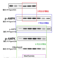
The right-side 3 bands (first, second and third bands from right) of IkB-alpha image, top-panel in Figure 4(A) of Ref.2 are similar to the second, third and fourth bands from left of pAMPK image in Figure 1(A) of Ref.2.
Moreover, the first and second bands from left of IkB-alpha image, top-panel in Figure 4(A) of Ref.2 is similar to both the fifth and sixth bands from left of pAMPK image in Figure 1(A) of Ref.2 and the fifth and sixth bands from left of pAMPK image in Figure 1(a) of Ref.4..
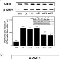
Furthermore, the pAMPK image (all 7 bands) in Figure 1(A) of Ref.2 is similar to the pAMPK image (all 7 bands) in Figure 1(a) of Ref.4 ,and first to sixth bands from left of the above pAMPK image of Ref.2,9 are similar to the pAMPK image for cilostazol in Figure 1(D) of Ref.2 and the pAMPK image in Figure 2(A) of Ref.1 and the pAMPK image for Metformin in Figure 3(b) of Ref.3.
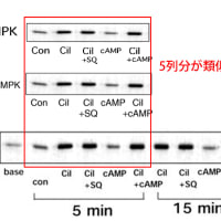
In addition, the rightside 5 bands (2.5 min 〓 60 min) of the pAMPK imgae in Figure 2(A) of Ref.1 are similar to upside-down and left-right reversal of the pAMPK image in Figure 3(A) of Ref.1.
__________________
References
1,
J Atheroscler Thromb. 2010;17(5):503-9. Epub 2010 Feb 24.
2,
Cardiovasc Res. 2009 Jan 1;81(1):133-9. Epub 2008 Aug 14.
3,
Hypertens Res. 2009 Sep;32(9):765-9. Epub 2009 Jul 10.
4.
Am J Hypertens. 2008 Apr;21(4):451-7. Epub 2008 Feb 7.
_____________________
Sincerely yours,
Juuichi Jigen
E-mail: 11jigen@mail.goo.ne.jp
Hypertension誌からの返信メール
件名
RE: Allegations of fabrication and falsification (Hypertension 2006 Jun;47(6):1183-8.)
送信者
John E. Hall <jehall@umc.edu>
宛先
'Juuichi Jigen' <11jigen@mail.goo.ne.jp> ; hypertension <hypertension@umc.edu>
CC
hypertension <hypertension@umc.edu>
日時
2011年02月07日 05:16:37
Dear Dr. Jigen,
We have reviewed the manuscript published in Hypertension (Hypertension 2006 Jun;47(6):1183-8.) as well as manuscripts 5 and 6 (referenced below) which you allege contain "similar" data/images.
We agree that the images appear to be similar, although not exactly the same. It is possible to have a "similar" image/data when one measures the same variable in different experiments.
We take charges of "fabricated" and "Falsified" data seriously. However, we will need additional information on why you believe the data published in Hypertension is "fabricated" or "falsified".
John E. Hall
Editor
-----Original Message-----
From: Juuichi Jigen [mailto:11jigen@mail.goo.ne.jp]
To: hypertension; John E. Hall
Cc: hypertension
Subject: Allegations of fabrication and falsification (Hypertension 2006 Jun;47(6):1183-8.)
Dear Dr.John E. Hall, Editor-in-Chief of Hypertension,
Please investigate fraudulent data of the article "Metformin inhibits Cytokine-Induced NF-kB Activation via AMPK Activation in Vascular Endothelial Cells" Hattori Y, Suzuki K, Hattori S, Kasai K., published in Hypertension (2006 Jun;47(6):1183-8.). You can find fabricated (falsified) Figure 3(A),(B),(C) and Figure 4(B) : namely,
(1)
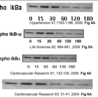
The Phospho-IkB-alpha image in Figure 4(A) of Ref.1 is similar to the Phospho-IkB-alpha image in Figure 5(A) of Ref.2 and the Phospho-IkBa image in Figure 3(A) of Ref.4 and the Phospho IkB-alpha image in Figure 6(A) of Ref.6.
(2)
The IkB-alpha image in Figure 4(B) of Ref.1 is similar to the IKB-alpha image in Figure 5(B) of Ref.2 and the IkBa image in Figure 3(B) of Ref.4 and the leftside 4 bands of the ONOO-IkB-alpha image in Figure 6(A) of Ref.6.
(3)
The IkB-alpha image in Figure 5(A) of Ref.2 is similar to both the IkB-a image in Figure 3(A) of Ref.4 and the IkB-alpha image in Figure 6(A) of Ref.6.
(4)
The right-side 3 bands (second, third and fourth bands from left) of IkB-alpha image, top-panel in Figure 3(C) of Ref.4 is similar to the middle 3 bands (second, third and fourth bands from left) of IkB-alpha image, top-panel in Figure 6(B) of Ref.6.
(5)
The IKKa/b image in Figure 3(C) of Ref.4 is similar to the left-side 4 bands (first to fourth bands from left) of the IKKalpha/beta image, fifth panel from bottom in Figure 6(B) of Ref.6.
Moreover, the left-side 3 bands of these two images are similar to the IKKalpha/beta image, second panel from bottom in Figure 6(B) of Ref.6..
(6)
The IkB-a image, bottom panel in Figure 3(C) of Ref.4 is similar to the left-side 4 bands (first to fourth bands from left) of IkB-alpha image, fourth panel from bottom in Figure 6(B) of Ref.6.
Moreover, the left-side 3 bands of these image are similar to the IkB-alpha image, bottom panel in Figure 6(B) of Ref.6.
(7)
The eNOS image in Figure 4(A) of Ref.3 is similar to the right-side 6 bands (for BH4 only, AngII, AngII/BH4) of eNOS Protein image in Figure 5(a) of Ref.5.
Moreover, these two images are similar to the 6 bands (third to eighth bands from left) of the MCP-1 image in Figure 4(B) of Ref.4.
__________________
References
1
Cardiovasc Res. 2009 Jan 1;81(1):133-9. Epub 2008 Aug 14.
2,
Life Sci. 2008 Apr 9;82(15-16):884-91. Epub 2008 Feb 16.
3,
Eur J Pharmacol. 2007 Jan 19;555(1):48-53. Epub 2006 Oct 18.
4,
Hypertension. 2006 Jun;47(6):1183-8. Epub 2006 Apr 24.
5,
J Hypertens. 2005 Jul;23(7):1375-82.
6,
Cardiovasc Res. 2004 Jul 1;63(1):31-40.
__________________
Sincerely yours,
Juuichi Jigen
E-mail: 11jigen@mail.goo.ne.jp
Cardiovascular Research誌からの返信メール
名
AW: Allegations of fabrication and falsification (Cardiovasc Research 2009 11:143-9)
送信者
CVR, Martinson <CVR@physiologie.med.uni-giessen.de>
宛先
Juuichi Jigen <11jigen@mail.goo.ne.jp>
日時
2011年02月07日 22:50:37
Dear Juuichi Jigen,
We have received your emails and our publisher has contacted us regarding this case as well. This is just to inform you that we take the accusations seriously and will investigate this situation ourselves. I will let you know what our Editorial Team's reply will be in a few days.
With kind regards,
Elizabeth Martinson
___________________________________
Elizabeth A. Martinson, Ph.D.
Managing Editor, Cardiovascular Research
Institute of Physiology
Aulweg 129
35392 Giessen, GERMANY
Tel. +49 (0)641 99 47 242
Fax +49 (0)641 99 47 209
Email CVR@physiologie.med.uni-giessen.de
-----Ursprüngliche Nachricht-----
Von: Juuichi Jigen [mailto:11jigen@mail.goo.ne.jp]
An: CVR, Martinson
Betreff: Allegations of fabrication and falsification (Cardiovasc Research 2009 11:143-9)
Dear Dr. Hans Michael Piper,
Please investigate fraudulent data of the article “Cilostazol inhibits cytokine-induced nuclear factor-kappaB activation via AMP-activated protein kinase activation in vascular endothelial cells.” Hattori Y, Suzuki K, Tomizawa A, Hirama N, Okayasu T, Hattori S, Satoh H, Akimoto K, Kasai K., published in Cardiovasc Research (2009 Feb;11(2):143-9). You can find fabricated (falsified) Figure 1(A),(B),(C),(D) and Figure4(A),(B),(C) : namely,
(1)
The first, second and third bands from right of IkB-alpha image, top-panel in Figure 4(A) of Ref.3 is similar to the second, third and fourth bands from left of pAMPK image in Figure 1(A) of Ref.3.
Moreover, the first and second bands from left of IkB-alpha image, top-panel in Figure 4(A) of Ref.3 is similar to both the fifth and sixth bands from left of pAMPK image in Figure 1(A) of Ref.3 and the fifth and sixth bands from left of pAMPK image in Figure 1(a) of Ref.7..
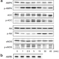
Furthermore, the pAMPK image (all 7 bands) in Figure 1(A) of Ref.3 is similar to the pAMPK image (all 7 bands) in Figure 1(a) of Ref.7 ,and first to sixth bands from left of the above pAMPK image of Ref.3,9 are similar to the pAMPK image for cilostazol in Figure 1(D) of Ref.3 and the pAMPK image in Figure 2(A) of Ref.2 and the pAMPK image for Metformin in Figure 3(b) of Ref.4.
In addition, the rightside 5 bands (2.5 min 〓 60 min) of the pAMPK imgae in Figure 2(A) of Ref.2 are similar to upside-down and left-right reversal of the pAMPK image in Figure 3(A) of Ref.2.
(2)
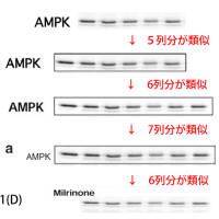
The AMPK image in Figure 1(A) of Ref.3 is similar to the APMK image in Figure 1(a) of Ref.7.
Moreover, the leftside 6 bands of these two APMK image are similar to the AMPK image in Figure 2(A) of Ref.2.
Furthermore, rightside 5 bands of this AMPK image in Figure 2(A) of Ref.2 are similar to the pAMPK image for Milrinone in Figure 1(D) of Ref.3
(3)
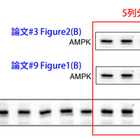
The rightside 5 bands of the AMPK image in Figure 1(B) of Ref.3 is similar to both the AMPK image in Figure 2(B) of Ref.2 and the AMPK image in Figure 1(b) of Ref.7.
Likewise, 5 bands (second to sixth bands from left) of pAMPK image in Figure 1(B) of Ref.3 are similar to the pAMPK image in Figure 2(B) of Ref.2 and the pAMPK image in Figure 1(b) of Ref.7.
(4)
The ACC and pACC images in Figure 1(A) of Ref.3 are similar to the ACC and pACC images in Figure 1(a) of Ref.7, respectively.
Moreover, the leftside 6 bands of these two images are similar to the ACC and p-ACC images in Figure 2(A) of Ref.2.
(5)
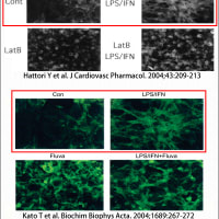
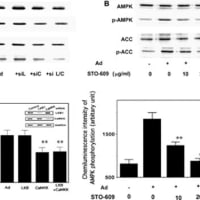
The Phospho-IkB-alpha image in Figure 4(A) of Ref.3 is similar to the Phospho-IkB-alpha image in Figure 5(A) of Ref.6 and the Phospho-IkBa image in Figure 3(A) of Ref.8 and the Phospho IkB-alpha image in Figure 6(A) of Ref.9.
(6)
The IkB-alpha image in Figure 4(B) of Ref.3 is similar to the IKB-alpha image in Figure 5(B) of Ref.6 and the IkBa image in Figure 3(B) of Ref.8 and the leftside 4 bands of the ONOO-IkB-alpha image in Figure 6(A) of Ref.9.
(7) The GST-Ikb-alpha image of top panel in Figure4(C) of Ref.1 is similar to both GST-Ikb-alpha image of top panel in Figure4(C) of Ref.3 and IkB-alpha image of top panel in Figure5(C) of Ref.6
(8) The IKKalpha/beta image in Figure 4(C) of Ref.3 is similar to both the ACC image in Figure 1(A) of Ref.6 and the IKKalpha/beta image in Figure 5(C) of Ref.6.
(9) The GST-Ikb-alpha image of bottom panel in Figure4(C) of Ref.3 is similar to GST-Ikb-alpha image of bottom panel in Figure4(C) of Ref.6. Moreover, these two images are similar to both the left-right reversal of the Akt image in Figure 1(A) of Ref.1 and the upside-down + left-right reversal of the GST-Ikb-alpha image of bottom panel in Figure4(C) of Ref.1
(10)
The GST-IkB-alpha image, bottom panel in Figure 4(C) of Ref.3 is similar to the Akt image in Figure 1(A) of Ref.6.
(11)
The left-side 4 bands (first to fourth bands from left) of the AMPK image in Figure 1(C) of Ref.3 is similar to the left-side 4 bands (first to fourth bands from left) of the AMPK image in Figure 2(A) of Ref.5.
(12)
The left-side 3 bands (first to third bands from left) of pAMPK image in Figure 1(C) of Ref.3 is similar to the left-side 3 bands (firtst to third bands from left) of pAPMK image in Figure 2(A) of Ref.5
__________________
References
1,
Metabolism. 2010 Jun 25. [Epub ahead of print] Fenofibrate suppresses microvascular inflammation and apoptosis through adenosine monophosphate-activated protein kinase activation.
Tomizawa A, Hattori Y, Inoue T, Hattori S, Kasai K.
2,
J Atheroscler Thromb. 2010;17(5):503-9.
3,
Cardiovasc Res. 2009 Jan 1;81(1):133-9.
4,
Hypertens Res. 2009 Sep;32(9):765-9.
5,
FEBS Lett. 2008 May 28;582(12):1719-24.
6,
Life Sci. 2008 Apr 9;82(15-16):884-91.
7,
Am J Hypertens. 2008 Apr;21(4):451-7.
8,
Hypertension. 2006 Jun;47(6):1183-8.
9,
Cardiovasc Res. 2004 Jul 1;63(1):31-40.
____
Sincerely yours,
Juuichi Jigen
E-mail: 11jigen@mail.goo.ne.jp


