| 論文番号 | 著者名 | 論文タイトル名 | 発表雑誌名 | 巻、号 | ページ | 出版年 |
| 論文#20 | Hattori Y, Kasai K. | Disruption of the actin cytoskeleton up-regulates iNOS expression in vascular smooth muscle cells. | J Cardiovasc Pharmacol. | 43(2) | 209-13 | 2004 |
| 論文#22 | Kato T, Hashikabe H, Iwata C, Akimoto K, Hattori Y. | Statin blocks Rho/Rho-kinase signalling and disrupts the actin cytoskeleton: relationship to enhancement of LPS-mediated nitric oxide synthesis in vrascular smooth muscle cells. | Biochim Biophys Acta. | 1689(3) | 267-272 | 2004 |
(A)- 指摘項目No.22
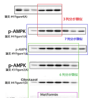
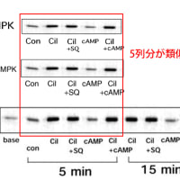
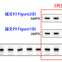
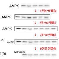
論文#20のFigure3と論文#22のFigure5について、二つの実験は、別々に行われた実験であるにも関わらず、画像が類似しているため、極めて不自然であり、データの流用の可能性と、少なくともどちらかの論文が捏造である可能性があります。(論文#20では薬剤との培養時間が1時間+LPS/IFNγとの培養時間が1時間の計2時間であり、一方、論文#22では、薬剤との培養時間が6時間+LPS/IFNγとの培養時間が1時間の計7時間です。つまり、両論文ではそもそも実験条件が全くことなるため、対照画像であるContの画像や、LPS/IFNの画像が類似していることは極めて不自然であり、画像を流用していると断言できます。)
論文撤回Watch様にて、詳しく、解説がなされています。
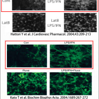
赤で囲んだ二つの図が同一であることが、明らかです。
上の図は論文#20のFigure3。下の図は論文#22のFigure5です。
画像の流用(データ捏造、改竄)が疑われます。

↓上記論文#20のFigure3

Hattori Y et. al. Journal of Cardiovascular Pharmacology. 43(2):209-213, February 2004.
Figure 3 Latrunculin B (Lat B) inhibits F-actin polymerization. VSMC were either untreated or treated with 0.5 [mu]M Lat B for 1 hour, and then either unstimulated or stimulated with LPS (30 [mu]g/ml) and interferon-[gamma] (10 ng/ml) for 1 hour. Cells were then fixed with 3.7% formaldehyde solution, washed with acetone, and stained with 165 nM NBD-phallacidin. Stained F-actin was imaged using a Nikon epifluorescent microscope.
J Cardiovasc Pharmacol. 2004 Feb;43(2):209-13.
↓上記論文#22のFigure5
Biochimica et Biophysica Acta 1689 (2004) 267– 272
Copyright 2004 Elsevier B.V. All rights reserved.
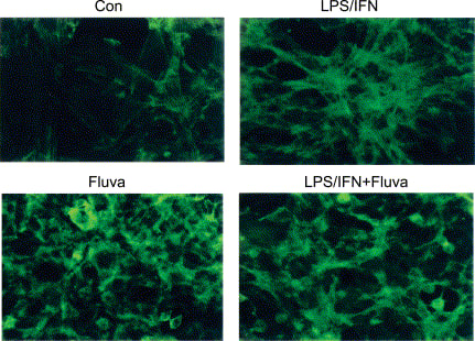
Fig. 5. Fluvastatin inhibits F-actin polymerization. VSMC were either untreated or treated with 25 AM fluvastatin for 6 h, after which they were either
unstimulated or stimulated with LPS (30 Ag/ml) and IFN (10 ng/ml) for 1 h. Cells were then fixed with a 3.7% formaldehyde solution, washed with acetone,
and stained with 165 nM NBD-phallacidin. Stained F-actin was imaged using a Nikon epifluorescent microscope.