| 論文番号 | 著者名 | 論文タイトル名 | 発表雑誌名 | 巻、号 | ページ | 出版年 |
| 論文#3 | Aoki C, Hattori Y, Tomizawa A, Jojima T, Kasai K. | Anti-inflammatory role of cilostazol in vascular smooth muscle cells in vitro and in vivo. | Journal of Atherosclerosis and Thrombosis |
17(5) | 503-509 | 2010 |
| 論文#4 | Hattori Y, Suzuki K, Tomizawa A, Hirama N, Okayasu T, Hattori S, Satoh H, Akimoto K, Kasai K. | Cilostazol inhibits cytokine-induced nuclear factor-kappaB activation via AMP-activated protein kinase activation in vascular endothelial cells. | Cardiovasc Research | 81(1) | 133-139 | 2009 |
| 論文#7 | Hattori Y, Nakano Y, Hattori S, Tomizawa A, Inukai K, Kasai K. | High molecular weight adiponectin activates AMPK and suppresses cytokine-induced NF-κB activation in vascular endothelial cells | FEBS Letters | 582(12) | 1719-1724 | 2008 |
| 論文#9 | Suzuki K, Uchida K, Nakanishi N, Hattori Y. | Cilostazol activates AMP-activated protein kinase and restores endothelial function in diabetes. | American Journal of Hypertension | 21(4) | 451-457 | 2008 |
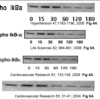
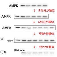
(A)- 指摘項目No.1
論文#4のFigure 4(A)の最上段のIkBαの画像の右3列分(左から4列目、5列目、6列目)の画像は、
同論文#4のFigure 1(A)のpAMPKの画像の左から2列目、3列目、4列目の画像と類似しています。
さらに、論文#4のFigure 4(A)の最上段のIkBαの画像(最上段の)の左から1列目、2列目の画像は、
同論文#4のFigure 1(A)のpAMPKの画像の左から5列目、6列目の画像や、
論文#9のFigure 1(a)のpAMPK画像の左から5列目、6列目の画像と、
類似しています(縦横比が改変されています。)
また、この論文#4のFigure1(A)のpAMPKの全7列分の画像は、
論文#9のFigure 1(a)のp-AMPKの全7列分の画像と類似しており、
また、これら#4,#9の二つの画像の左6列分と、
論文#4のFigure 1(D)のpAMPKのCilostazolの画像全6列分や、
論文#3のFigure 2(A)のpAMPKの画像全6列分や、
論文#5のFigure 3(b)のMetformin pAMPKの画像全6列分とも
類似していることから、データの捏造(流用)が疑われます。
また、この論文#3のFigure 2(A)のpAMPKの画像の右5列分(2.5 min~60 minの画像)を、
上下左右逆にしたものが、同論文#3のFigure 3 (A)のpAMPKの画像に類似しています。
以上より、データの捏造(流用)が疑われます。
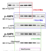
(A)- 指摘項目No.2
論文#4のFigure 1(A)のAMPKの画像の全7列分は、
論文#9のFigure 1(a)のAMPKの画像の全7列分と類似しています。
さらに、この論文#4と#9の画像のうち、左6列分(左から1,2,3,4,5,6列目)が、
論文#3のFigure 2(A)のAMPKの画像に類似しています。
さらに、この論文#3のFigure 2(A)のAMPKの画像6列分のうち、右5列分が、
同論文#3のFigure 3(A)のAMPKの画像に類似しています。
さらに、上記の論文#4と#9の画像のうち、右6列分(左から2,3,4,5,6,7列目)が、
論文#4のFigure 1(D)のpAMPKのMilrinoneの画像に類似しています(コントラストと縦横比を変更しています。)
以上より、データの捏造(流用)が疑われます。
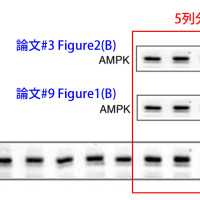
(A)- 指摘項目No.3-a
論文#4のFigure 1(B)のAMPKの画像10列分のうち、右側5列分が、
論文#3のFigure 2(B)のAMPKの画像全5列分や、
論文#9のFigure 1(b)のAMPKの画像全5列分に、類似しています(縦横比が変更されてます。)。
(A)- 指摘項目No.3-b
同様に、
論文#4のFigure 1(B)のpAMPKの画像10列分のうち、左から2,3,4,5,6,列目の5列分が、
論文#3のFigure 2(B)のpAMPKの画像全5列分や、
論文#9のFigure 1(b)のpAMPKの画像全5列分に、
類似しており、データの捏造(流用)が疑われます(縦横比が変更されてます。)。
(A)- 指摘項目No.4
さらに、
論文#4のFigure 1(A)のACC画像と、pACC画像の全7列分は、
論文#9のFigure 1(a)のACC画像と、pACC画像 の全7列分と、類似しています。
さらに、これら#4, #9の画像の左側6列分の画像が、
論文#3のFigure 2(A)のACC画像と、p-ACC画像の全6列分と、類似しており、
データの捏造(流用)が疑われます。
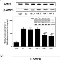
(A)- 指摘項目No.19
論文#4のFigure 1(C)のAMPKの画像の左4列分(左から1、2、3、4列目)の画像は、
論文#7のFigure 2(A)のAMPKの画像の左4列分(左から1、2、3、4列目)の画像と
類似しており(縦横の比率が変更されています)、データの捏造(流用)が疑われます。
(A)- 指摘項目No.20
論文#4のFigure 1(C)のpAMPKの画像の左3列分(左から1、2、3列目)の画像は、
論文#7のFigure 2(A)のpAMPKの画像の左3列分(左から1、2、3列目)の画像と
類似しており(縦横の比率が変更されています)、データの捏造(流用)が疑われます。
↓論文#3のFigure 2
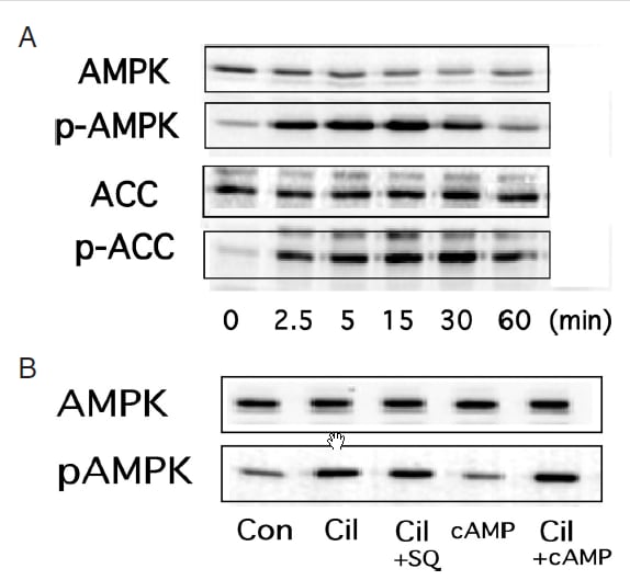
Journal of Atherosclerosis and Thrombosis Vol. 17 (2010) , No. 5 503-509
Copyright (c) 2010 Japan Atherosclerosis Society
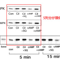
(A) Cilostazol-mediated activation of AMPK in rat VSMC. VSMC were treated with colostazol (100μM) for the indicated time periods before lysis, after which samples of cell lysate were probed with antibodies specific for the phosphorylated forms of AMPK and acetyl-CoA carboxylase (ACC). (B) HUVEC treated with cilostazol (100 μM) alone or in the presence of an adenylate cyclase inhibitor SQ22536 (10 μM) or a cell-permeable cAMP analog pCTP-cAMP (100 μM). After 15 min of incubation, the cells were lysed and p-AMPK activity was analyzed. Three independent studies showed similar results.
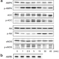
↓論文#4のFigure 1(画像をクリックすると解像度の高い画像が表示されます。)
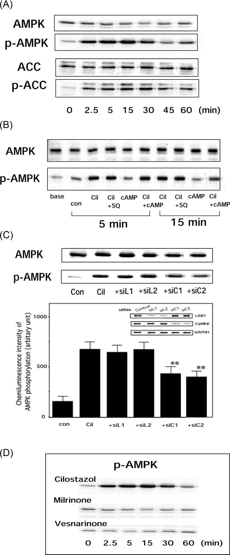
Copyright © 2011 European Society of Cardiology
Cardiovasc Res (2009) 81 (1): 133-139.
Figure 1
(A) Cilostazol activates AMP-activated protein kinase (AMPK) in vascular endothelial cells. Human umbilical vein endothelial cells (HUVEC) were treated with cilostazol (100 μM) for the indicated time periods before lysis, after which each cell lysate sample was probed with antibodies specific for phosphorylated forms of AMPK and acetyl-CoA carboxylase (ACC). (B) HUVEC were treated with cilostazol (100 μM) alone or in the presence of an adenylate cyclase inhibitor SQ 22536 (10 μM) or a cell-permeable cyclic AMP (cAMP) analogue pCTP-cAMP (100 μM). After 5 and 15 min of incubation, the cells were lysed and p-AMPK was analysed. Three independent studies showed similar results. (C) Cilostazol activates AMPK, which was significantly attenuated in HUVEC transfected with CaMKKβ siRNA (siC1 or siC2: 10 nM) but not with LKB1 siRNA (siL1 or siL2: 10 nM). Inset (lower figure): 48 h after cells were transfected with control siRNA, siL1, siL2, siC1, or siC2, the mRNA levels of LKB1, CaMKKβ, and GAPDH were determined. (D) Cilostazol, but not milrinone or vesnarinone, activates AMPK in vascular endothelial cells. HUVEC were treated with cilostazol (100 μM), milrinone (100 μM), or vesnarinone (100 μM) for the indicated time periods before lysis, after which each cell lysate sample was probed with antibodies specific for phosphorylated AMPK.
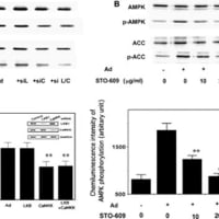
↓論文#7のFigure 2(画像をクリックすると解像度の高い画像が表示されます。)

Copyright © 2008 Published by Elsevier B.V.
FEBS Letters Volume 582, Issue 12, 28 May 2008, Pages 1719-1724
Fig. 2. (A) HMW adiponectin (Ad) activates AMPK, which was significantly attenuated in HUVEC transfected with CaMKKβ siRNA (siC: 10 nM) but not with LKB1 siRNA (siL: 10 nM). Inset (lower figure): 48 h after cells were transfected with control, LKB1, or CaMKKβ siRNA, the mRNA levels of LKB1 or CaMKKβ were determined. (B) HMW adiponectin (Ad)-induced AMPK phosphorylation was inhibited by the inhibitor of CaMKKβ, STO-609 in HUVEC. Results represent the means ± S.D. (n = 4). **P < 0.01 vs. AMPK activity by Ad.
↓論文#9のFigure 1
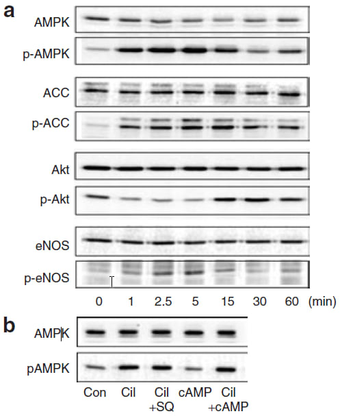
© 2011 American Journal of Hypertension, Ltd.l
American Journal of Hypertension 21, 451-457 (April 2008)
Figure 1: Cilostazol activates AMP-activated protein kinase (AMPK) in
vascular endothelial cells. (a) human umbilical vein endothelial cells (HUVECs)
were treated with 100 μmol/l cilostazol for the indicated time periods before
lysis, after which cell lysates were probed with antibodies specific for AMPK,
acetyl-CoA carboxylase (ACC), Akt, or endothelial nitric oxide synthase
(eNOS), or their phosphorylated forms.(b)HUVEC were treated with cilostazol (100 μmol/l) alone or in the presence of an adenylate cyclase inhibitor SQ22536 or a cell-permeable cAMP analog pCTP-cAMP. After 5-min incubation, cells were lysed and p-AMPK was analysed.