獨協医科大学の研究不正問題

獨協医科大学内分泌代謝内科研究室における 論文捏造・二重投稿の疑惑追及ブログ

続 獨協内分泌代謝内科Diabetologia

2011年01月24日 | 論文撤回Watch


written by Retraction Watch / 2011.06.18 09:07


続 獨協内分泌代謝内科Diabetologia

獨協医大内分泌代謝内科Diabetologiaの続きです。

Dr次元さまよりのコメントによると

積極的に対処しようとしてくれたのは、Diabetologia誌だけでした

とのことですので、 Diabetologiaに掲載されている別の論文を眺めてみました。


[画像: ]


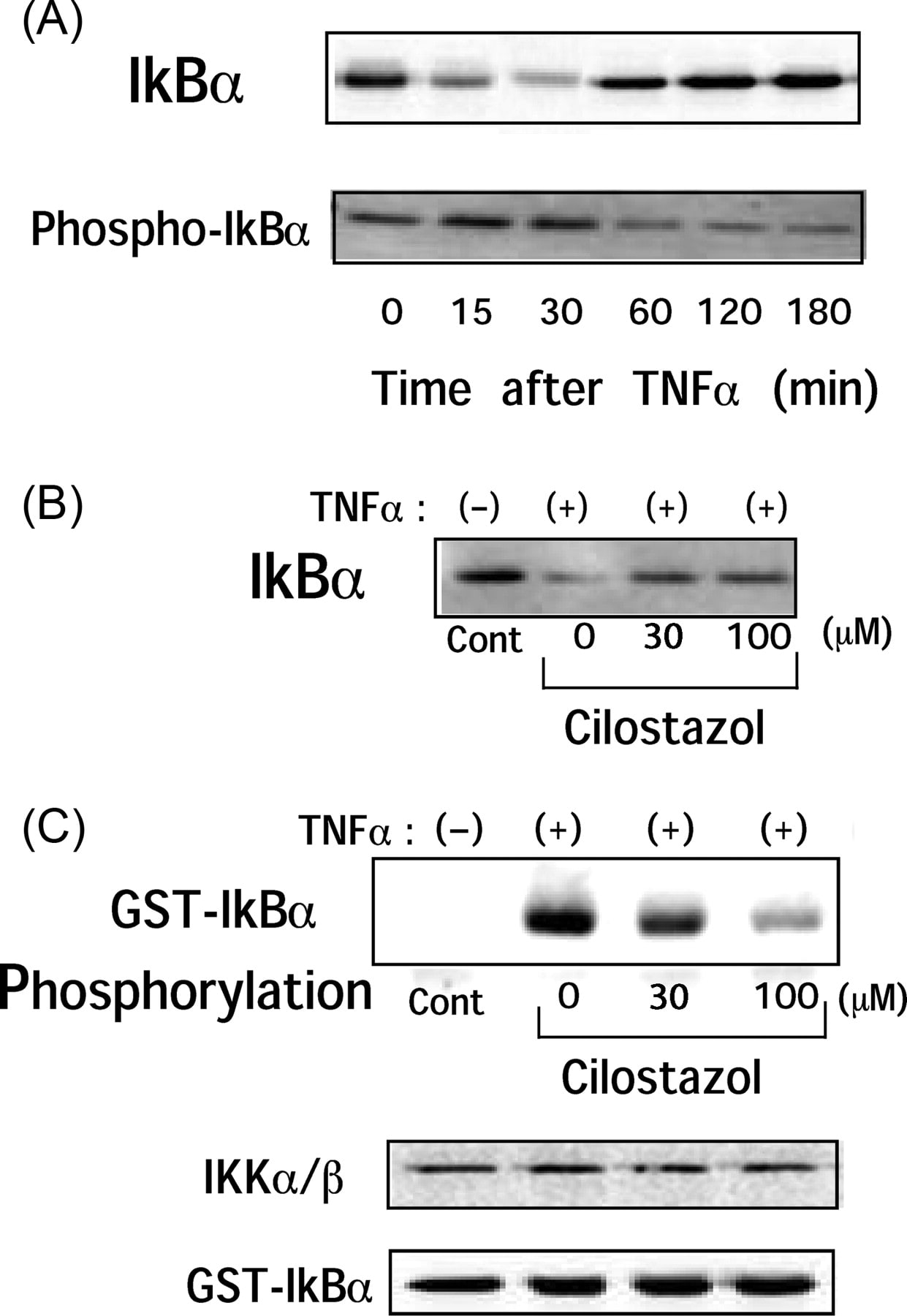
図2Cを左右に反転して縦横比をいじると、別の論文の図に似通っているように思いました。 


written by Retraction Watch / 2011.06.18 09:07


獨協内分泌代謝の撤回4論文

2011年01月24日 | 論文撤回Watch

獨協内分泌代謝の撤回4論文

Retraction Watch / 2011.08.26 09:26 / 推薦数 : 1

獨協医大内分泌代謝内科の撤回された4本の論文について

詳しく掘り下げます。


まず、最初のペアー

Diabetologia論文

Metabolism論文

を比べます。


Diabetologia論文

の論文受理日は、2004年11月9日

掲載許可日は、2005年2月8日

オンライン発表が 2005年4月30日

論文撤回発表が、2011年5月17日


Metabolism論文

の論文受理日は、2004年8月4日

掲載許可日は、2004年10月25日

オンライン発表が 2005年3月23日

論文撤回発表が、2011年6月15日


責任著者は両論文とも同一人物です。 


責任著者はMetabolism論文掲載許可の通知を受けた後

Diabetologia論文

を投稿した可能性が高いと考えられます。


Metabolism論文の著者は3名で

すべて獨協医大内分泌代謝内科


一方 Diabetologia論文の著者は

上記3人に加えて、

獨協医大分子生物学1名と

米国コーネル大学の1名、計5名。


Diabetologia論文の撤回は、編集長の判断によるもの、

(すなわち、著者らの要求によるものとの記載はないし、また著者らが撤回に同意したかどうかも記載はない)

一方

Metabolism論文の撤回は、責任著者の要求によるもの

となっています。


Metabolism論文の撤回理由は明白に記載されていません。

This article contains material that has appeared in Diabetologia, 48 (2005) 1066–1074.


と記載されていますが、

仮に、

Diabetologia論文にだけ、データ改ざん等の不正、

あるいは、図の再利用があり、かつ

Metabolism論文には、何ら問題なし、

となると、

Metabolism論文は先行論文でもあるため、

撤回する理由は、ないことになります。


責任著者が、撤回するに値する何らかの理由があることを

認めていると判断するのが

妥当かと考えます。 


8月も残すところわずか。


獨協医大の調査委員会は7,8月ごろには調査結果を公表する、とされていましたが、どうなっているのかしら。


獨協医大内分泌代謝内科 二つ目のペアー

2011年01月24日 | 論文撤回Watch


獨協医大内分泌代謝内科 二つ目のペアー

Retraction Watch / 2011.08.27 00:18 / 推薦数 : 1

獨協医大内分泌代謝内科の撤回された4本の論文について

詳しく掘り下げます。


今回は二つ目のペアー

JCP 論文

BBA 論文

を比べます。


JCP 論文

の論文受理日は、2003年6月27日

掲載許可日は、2003年9月8日


BBA 論文

の論文受理日は、2004年2月3日

掲載許可日は、2004年4月21日

オンライン発表が 2004年5月31日


両論文撤回発表が、2011年7月6日


責任著者は両論文とも同一人物です。 


JCP 論文の著者はたった2名で獨協医大内分泌代謝内科


一方 BBA 論文の著者は5名で、JCP論文の責任著者でない方の1名は含まれていません。


さてこの両論文で問題となっているのは、1点のみ。

BBA論文の図5の一部が、JCP論文の図3の一部と同一であること(ありえないのに)。図を再掲します。


両雑誌の編集長が、責任著者に連絡をとり説明を求めましたが、結局十分な回答は、得られず、編集長の判断で両論文とも撤回となりました。


さて、

ここで獨協医大の問題から離れます。


Dr 次元さまのとりあげている

この 二つの図を見比べてください。

明らかに同じ顕微鏡写真を、条件の違う別々の実験結果として、発表しているわけであり、

今回とりあげた獨協医大の例と本質的には同じです。

従って、これらの論文2本とも撤回されても

不思議ではありません。


大将の座を獲得できたかもしれませんが、

舞台はスウェーデンのようだったので、

米国研究不正取り締まり局の管轄外

となり、助かりました。


獨協医大内分泌代謝内科 震災の影響を考慮した編集長

2011年01月24日 | 論文撤回Watch

written by Retraction Watch / 2011.08.24 11:26


獨協医大内分泌代謝内科 震災の影響を考慮した編集長

獨協医大内分泌代謝内科の研究不正問題の続報です。


今回2本の論文が撤回されました。


[画像: ]


2本の論文は別々の実験なのに、なぜか

同じ顕微鏡写真を使って

論文発表しています。


両雑誌の編集長がお互い相談して、両方とも撤回することに決めたようです。


編集長は、著者らに説明を求めました。

当初、著者らから返答があったが、震災後に返答がこなくなったとのことです。


ひとつの論文は生き残れる可能性はあったのですが、

2本とも撤回になったのは、当然の成り行きだと

思います。


これで、

獨協医大内分泌代謝内科からの一連の論文撤回は

4本となりました。


1本目

以前に紹介しました。

これに対応する形で

別の論文も撤回されました。

両論文で同じ図を使っていたという、同じ手口です。


詳しくは

Dr 次元さまのサイトへ


おそらく4本の撤回は

Dr次元さまからの雑誌社への研究不正疑いの連絡が

きっかけになったと思われます。

written by Retraction Watch / 2011.08.24 11:26


3本一気に!獨協医大

2011年01月24日 | 論文撤回Watch

written by Retraction Watch / 2011.09.18 00:54


3本一気に!獨協医大

獨協医大代謝内分泌内科の研究グループからの論文3本

が一気に撤回されました。


雑誌Cardiovasc Res 最新号に撤回のアナウンスがなされました。


3本の撤回論文は、

Cardiovasc Res. 2009;81(1):133-9.

Cilostazol inhibits cytokine-induced nuclear factor-kappaB activation via AMP-activated protein kinaseactivation in vascular endothelial cells

Hattori Y, Suzuki K, Tomizawa A, Hirama N, Okayasu T, Hattori S, Satoh H, Akimoto K, Kasai K.

Source

Department of Endocrinology and Metabolism, Dokkyo University School of Medicine

Cardiovasc Res. 2004;63(1):31-40.

NO suppresses while peroxynitrite sustains NF-kappaB:

paradigm to rationalize cytoprotective andcytotoxic 

actions attributed to NO

Hattori Y, Kasai K, Gross SS.

Source

Department of Endocrinology and Metabolism, Dokkyo University School of Medicine

Cardiovasc Res. 2002;54(3):649-58.

Statin enhances cytokine-mediated induction of nitric oxide synthesis in vascular smooth muscle cells.

Hattori Y, Nakanishi N, Kasai K.

Source

Department of Endocrinology and Metabolism, Dokkyo University School of Medicine 


3本とも原論文は無料で読めます。


3本とも撤回は雑誌社によりなされました。すなわち、著者らの要求による撤回ではないと、解釈できます。

明らかにデタラメ、インチキな論文は、著者の同意がなかっても雑誌社は撤回できるのです。


また詳しい解説は、Dr次元さまのサイトで読めます。

2009年論文の解説はこちら

2004年論文の解説はこちら

2002年論文の解説はこちら


これで 獨協医大 代謝内分泌からの一連の撤回論文は

合計7本となりました。


written by Retraction Watch / 2011.09.18 00:54


Dr次元さまよりコメント 獨協医大

2011年01月24日 | 論文撤回Watch


Dr次元さまよりコメント 獨協医大

written by Retraction Watch / 2011.10.17 01:40 


久しぶりにDr次元さまより

コメントを頂ました。


Dr次元さまの警笛が功を奏し、

Diabetologiaに掲載された獨協医大の論文が撤回になる予定とのことです。


以下7月14日に Dr次元さまが

Diabetologia編集長のDr Naylor

に宛てたメールの一部


Dear Dr J Naylor 
 
Recently, I found that you retracted Dr. Hattori's paper "Diabetologia. 2005 Jun;48(6):1066-74.". 

In addition, I found another image manupulation by Dr. Hattori in another Diabetologia paper ( 2010;53:2256-63.). 

Please compare Figure 2C image in the above Diabetologia paper (2010) and Figure 1B in the Cardiovascular Research paper.
Reversed image of band image in Figure 2C in Diabetologia paper (2010) is very similar to the band image in Figure 1B in Cardiovasc Res. paper (2009). 
 
I hope you find the attached jpg file on the image manupulation informative


そしてすぐに返事がくる。 


Many thanks for your email of 7th July. We are in fact already looking into the two papers you mention, and I'll let you know the outcome of our investigation


すでに検証は始めているとのこと。Dr次元さま以外に先に警笛を鳴らした人がいたのでしょうか。


そして今回、撤回にもっていくという返事。


Dear Dr Jigen 

I just wanted to update you with regards to the article published by Hattori et al in 2010. The concerns you raised were confirmed by an independent adviser to the journal. We gave Dr Hattori several opportunities to respond to these concerns, but heard nothing. We will now proceed with retracting the Diabetologia article. 

Many thanks for bringing this matter to our attention. 

Kind regards 

Judy 


撤回になる予定の論文は

こちら

Diabetologia. 2010 Oct;53(10):2256-63.  A glucagon-like peptide-1 (GLP-1) analogue, liraglutide, upregulates nitric oxide production and exerts anti-inflammatory action in endothelial cells.

Hattori Y, Jojima T, Tomizawa A, et al,


なにが問題だったかというと


[画像: ]


この図は以前に論文撤回Watchで紹介しました。


Dr次元さまの多くのサイトがこのところ

全く更新されていないので

心配しておりました。


今回、コメントを頂いたことで少し安堵しましたが、

Dr次元さまには、今後も益々ご活躍して頂きたいと

思っております。


written by Retraction Watch / 2011.10.17 01:40


誠意と節度をもった医療

2011年01月24日 | 論文撤回Watch


誠意と節度をもった医療

Retraction Watch / 2011.12.20 00:19 / 推薦数 : 2

「誠意と節度をもった医療を」

これは獨協医大内分泌代謝内科のモットーのようです。

R


ひさしぶりに獨協医大内分泌代謝内科です。


撤回された論文は

Diabetologia. 2010 Oct;53(10):2256-63. Epub 2010 Jul 1.

A glucagon-like peptide-1 (GLP-1) analogue, liraglutide, upregulates nitric oxide production and exerts anti-inflammatory action in endothelial cells.

Hattori Y, Jojima T, Tomizawa A, Satoh H, Hattori S, Kasai K, Hayashi T.

Source

Department of Endocrinology and Metabolism, Dokkyo University School of Medicine, Mibu, Tochigi 321-0293, Japan. yhattori@dokkyomed.ac.jp


 撤回のアナウンスです。


This article has been retracted by the Editor-in-Chief of Diabetologia following the discovery of redundant publication (part of Figure 2c was previously published in Cardiovascular Research)


この説明を補足する意味で 再度、問題の図を紹介します。

撤回のアナウンスに「左右反転」という説明があれば、

ここで図を紹介する必要はなかったのですが、、、 


Dr次元さまを含めた一般の読者が

雑誌社に対して警笛を鳴らした

ことが撤回の引き金となっています。


警笛を鳴らした方と

それに応じたDiabetologiaの編集部

に敬意を表します。


論文#1

2011年01月24日 | 論文捏造
論文番号 著者名 論文タイトル名 発表雑誌名 巻、号 ページ 出版年
論文#1 Aoki C, Nakano A, Tanaka S, Yanagi K, Ohta S, Jojima T, Kasai K, Takekawa H, Hirata K, Hattori Y. Fluvastatin upregulates endothelial nitric oxide synthase activity via enhancement of its phosphorylation and expression and via an increase in tetrahydrobiopterin in vascular endothelial cells. Int J Cardiol. [Epub ahead of print] [Epub ahead of print] 2011

論文番号
指摘項目番号

不正が疑われる図の番号


論文#1 指摘項目No.35
指摘項目No.36
Figure 1(A)
Figure 1(B)
論文1のFigure 1(A)の上から2段目のp-Ser1177 eNOSの画像の右5列分の画像は、
同論文1のFigure 1(A)の上から3段目のp-Ser633 eNOSの画像の右5列分の画像と、
類似しており、さらに、どちらの段の画像についても、左から1列目の画像と、その他5列分の画像が連続しておらず、つぎはぎされた編集の痕跡が確認できることから、
画像の流用(データの捏造)が疑われます。
論文1のFigure 1(B)の左上のeNOSの画像と、右上のeNOSの画像が
類似しており、画像の流用が疑われます。


研究不正に関連する図(Figure)

↓上記論文#1のFigure 1 (画像クリックで高解像度の画像を表示できます。)


© 2010 Elsevier Ireland Ltd. All rights reserved.
International Journal of Cardiology (2010)
Fig. 1. (A) Fluvastatin-stimulated endothelial nitric oxide (NO) synthase (eNOS) phosphorylation at Ser-1177 and Ser-633 in HUVEC. HUVEC were treated with fluvastatin (1 μM) for
the indicated times, and cells were lysed, and then immunoblotted with anti-phospho-Ser-1177 antibody and anti-phospho-Ser-1177 antibody and nonphosphospecific anti-eNOS
antibody. (B) Cells were pretreated for 30 min with 100 nM wortmannin (WT) or with 500 nM KT-5720 (KT) and were treated with fluvastatin (1 μM) for 15 min and
immunoblotted. Similar results were obtained in three independent experiments.


上記論文1のFigure 1(A)の上から2段目のp-Ser1177 eNOSの画像の右5列分の画像は、
同論文1のFigure 1(A)の上から3段目のp-Ser633 eNOSの画像の右5列分の画像と、
類似しており、さらに、どちらの段の画像についても、左から1列目の画像と、その他5列分の画像が連続しておらずつぎはぎされたものであることから、
画像の流用(データの捏造)が疑われる。


また、上記論文1のFigure 1(B)の左上のeNOSの画像と、右上のeNOSの画像が類似しており、
画像の流用が疑われる。


論文#2

2011年01月24日 | 論文捏造


論文番号 著者名 論文タイトル名 発表雑誌名 巻、号 ページ 出版年
論文#2 Tomizawa A, Hattori Y, Inoue T, Hattori S, Kasai K.

Fenofibrate suppresses microvascular inflammation and apoptosis through adenosine monophosphate-activated protein kinase activation.

Metabolism Clinical and Experimental [Epub ahead of print] [Epub ahead of print]

2010


論文番号
指摘項目番号

不正が疑われる図の番号


論文#2 指摘項目No.8
指摘項目No.10
指摘項目No.29
指摘項目No.30
指摘項目No.31
指摘項目No.32
指摘項目No.33
指摘項目No.34

Figure 4(C)
Figure 1(A), 4(C)
Figure 1(A)
Figure 1(A)
Figure 1(A)
Figure 4(A),(C)
Figure 4(A),(B)
Figure 4(B)

(A)- 指摘項目No.8
論文#2のFigure 4(C)の最上段のGST-IkB-αの画像は、
論文#4のFigure 4(C)の最上段GST-IkBαの画像や、
論文#8のFigure 5(C)の最上段のIkB-αの画像と、
類似しており、画像の流用(データの捏造)が疑われます。


(A)- 指摘項目No.10
論文#4のFigure 4(C)の最下段のGST-IkBαの画像と、
論文#8のFgure 5(C)の最下段のIkB-αの画像が、
類似しており、データの捏造(流用)が疑われます。
さらに、この二つの画像は、
論文#2のFigure 1Aの上から5段目のAktの画像を左右逆にした画像や、
同論文#2のFigure 4Cの最下段のGST-IkB-αの画像を上下左右逆にした画像とも、
類似しており、画像の流用(データの捏造)が疑われます。


(A)- 指摘項目No.29
論文#2のFigure 1Aの上から1段目と2段目のAMPKとpAMPKの画像は、
論文#8のFigure 1(A)の上から1段目と2段目のAMPKとpAMPKの画像と、
類似しており、画像の流用(データの捏造)が疑われます。


(A)- 指摘項目No.30
論文#2のFigure 1Aの上から6段目のp-Aktの画像は、
論文#8のFigure 1(A)の上から6段目のp-Aktの画像と、
類似しており、画像の流用(データの捏造)が疑われます。


(A)- 指摘項目No.31
論文#2のFigure 1Aの上から7段目のeNOSの画像は、
論文#8のFigure 1(A)の上から7段目のeNOSの画像を左右逆に加工した画像と同一であり、
画像の流用(データの捏造)が疑われます。


(A)- 指摘項目No.32
論文#2のFigure 4Aの最上段のIkB-αの一番左の1列分の画像(0 minの画像)は、
同論文#2のFigure 4Cの最下段のGST-IkB-αの一番左の1列分の画像(AGE-の画像)の縦横比を変更したものに、
類似しており、画像の流用(データの捏造)が疑われます。


(A)- 指摘項目No.33
論文#2のFigure 4Aの最上段のIkB-αの左から2,3,4列目の3列分は、
同論文#2のFigure 4BのIkB-αの画像の右3列分(Fenofibrate 0, 30, 100 μMの画像)の画像と類似しています(画像サイズを加工しています)。
さらに、この3列分の画像は、
論文#6のFigure 2(B)の最上段のp-Aktの画像の左3列分(0, 5, 15 minの画像)と
類似しており、画像の流用(データの捏造)が疑われます。


(A)- 指摘項目No.34
論文#2のFigure 4BのIkB-αの画像の一番左の列の画像(Contの画像)は、
論文#6のFigure 2(B)の一番上のp-Aktの画像の左から4列目の画像(30 minの画像)と
類似しており、画像の流用(データの捏造)が疑われます。


研究不正に関連する図(Figure)


↓上記論文#2のFigure 1


© 2010 Elsevier Inc. All rights reserved.
Metabolism Clinical and Experimental xx (2010)
Fig. 1. A, Fenofibrate activates AMPK in microvascular endothelial cells.
Human glomerular microvascular endothelial cells were treated with
100 μmol/L fenofibrate for the indicated periods before lysis, after which
cell lysates were probed with antibodies specific for AMPK, ACC, Akt,
or eNOS, or their phosphorylated forms. B, Peroxisome proliferator–
activated receptor α antagonist MK886 did not affect fenofibrateinduced
AMPK activation, and PPARα agonist WY14643 scarcely
activated AMPK in HGMEC. C, Fenofibrate activates AMPK in
microvascular endothelial cells but not in hepatic cells. HepG2 cells
and HGMEC were treated with fenofibrate, WY14643, or bezafibrate for
15 minutes and analyzed with antibodies specific for AMPK or ACC.
Lower figure in Fig. 1C shows that fenofibrate did not activate AMPK
during indicated period in HepG2. Fe indicates fenofibrate (100 μmol/L);
MK, MK886 (100 μmol/L); WY, WY14643 (100 μmol/L); Be,
bezafibrate (100 μmol/L).


↓上記論文#2のFigure 4


© 2010 Elsevier Inc. All rights reserved.
Metabolism Clinical and Experimental xx (2010)
Fig. 4. A, Human glomerular microvascular endothelial cells were incubated
with AGE-BSA for 0 to 180 minutes. The cells were lysed and subjected to
Western blot analysis using anti-IκBα and anti–phospho-IκBα antibodies.
B, The effect of fenofibrate on IκB-α degradation in human umbilical vein
endothelial cells. Cells were incubated for 30 minutes with fenofibrate (30
and 100 μmol/L), followed by AGE-BSA for 15 minutes. Cells were then
lysed and subjected to Western blot analysis using anti-IκBα antibody. C,
The effect of fenofibrate on IKK activity in HGMEC. Cells were incubated
for 30 minutes with fenofibrate (30 and 100 μmol/L), followed by AGEBSA
for 15 minutes. Cells were then lysed and immunoprecipitated with
anti-IKKα/β antibody and used for kinase assay using recombinant IκB-α as
a substrate. Note that equal band densities for IKKα/β- and IκB-α were
observed. Similar results in 3 independent experiments were obtained, and
representative photographs are shown.


論文#3

2011年01月24日 | 論文捏造
論文番号 著者名 論文タイトル名 発表雑誌名 巻、号 ページ 出版年
論文#3 Aoki C, Hattori Y, Tomizawa A, Jojima T, Kasai K. Anti-inflammatory role of cilostazol in vascular smooth muscle cells in vitro and in vivo. Journal of Atherosclerosis and Thrombosis 17(5) 503-509 2010

論文番号
指摘項目番号

不正が疑われる図の番号


論文#3 指摘項目No.1
指摘項目No.2
指摘項目No.3-a,b
指摘項目No.4
Figure 2(A), 3(A)
Figure 2(A), 3(A)
Figure 2(B)
Figure 2(A)


(A)- 指摘項目No.1
論文#4のFigure 4(A)の最上段のIkBαの画像の右3列分(左から4列目、5列目、6列目)の画像は、
同論文#4のFigure 1(A)のpAMPKの画像の左から2列目、3列目、4列目の画像と類似しています。
さらに、論文#4のFigure 4(A)の最上段のIkBαの画像(最上段の)の左から1列目、2列目の画像は、
同論文#4のFigure 1(A)のpAMPKの画像の左から5列目、6列目の画像や、
論文#9のFigure 1(a)のpAMPK画像の左から5列目、6列目の画像と、
類似しています(縦横比が改変されています。)
また、この論文#4のFigure1(A)のpAMPKの全7列分の画像は、
論文#9のFigure 1(a)のp-AMPKの全7列分の画像と類似しており、
また、これら#4,#9の二つの画像の左6列分と、
論文#4のFigure 1(D)のpAMPKのCilostazolの画像全6列分や、
論文#3のFigure 2(A)のpAMPKの画像全6列分や、
論文#5のFigure 3(b)のMetformin pAMPKの画像全6列分とも
類似していることから、データの捏造(流用)が疑われます。
また、この論文#3のFigure 2(A)のpAMPKの画像の右5列分(2.5 min~60 minの画像)を、
上下左右逆にしたものが、同論文#3のFigure 3 (A)のpAMPKの画像に類似しています。
以上より、データの捏造(流用)が疑われます。


(A)- 指摘項目No.2
論文#4のFigure 1(A)のAMPKの画像の全7列分は、
論文#9のFigure 1(a)のAMPKの画像の全7列分と類似しています。
さらに、この論文#4と#9の画像のうち、左6列分(左から1,2,3,4,5,6列目)が、
論文#3のFigure 2(A)のAMPKの画像に類似しています。
さらに、この論文#3のFigure 2(A)のAMPKの画像6列分のうち、右5列分が、
同論文#3のFigure 3(A)のAMPKの画像に類似しています。
さらに、上記の論文#4と#9の画像のうち、右6列分(左から2,3,4,5,6,7列目)が、
論文#4のFigure 1(D)のpAMPKのMilrinoneの画像に類似しています(コントラストと縦横比を変更しています。)
以上より、データの捏造(流用)が疑われます。


(A)- 指摘項目No.3-a
論文#4のFigure 1(B)のAMPKの画像10列分のうち、右側5列分が、
論文#3のFigure 2(B)のAMPKの画像全5列分や、
論文#9のFigure 1(b)のAMPKの画像全5列分に、類似しています(縦横比が変更されてます。)。
(A)- 指摘項目No.3-b
同様に、
論文#4のFigure 1(B)のpAMPKの画像10列分のうち、左から2,3,4,5,6,列目の5列分が、
論文#3のFigure 2(B)のpAMPKの画像全5列分や、
論文#9のFigure 1(b)のpAMPKの画像全5列分に、
類似しており、データの捏造(流用)が疑われます(縦横比が変更されてます。)。


(A)- 指摘項目No.4
さらに、
論文#4のFigure 1(A)のACC画像と、pACC画像の全7列分は、
論文#9のFigure 1(a)のACC画像と、pACC画像 の全7列分と、類似しています。
さらに、これら#4, #9の画像の左側6列分の画像が、
論文#3のFigure 2(A)のACC画像と、p-ACC画像の全6列分と、類似しており、
データの捏造(流用)が疑われます。


研究不正に関連する図(Figure)

↓論文#3のFigure 2


Journal of Atherosclerosis and Thrombosis
Vol. 17 (2010) , No. 5 503-509
Copyright (c) 2010 Japan Atherosclerosis Society
(A) Cilostazol-mediated activation of AMPK in rat VSMC. VSMC were treated with colostazol (100μM) for the indicated time periods before lysis, after which samples of cell lysate were probed with antibodies specific for the phosphorylated forms of AMPK and acetyl-CoA carboxylase (ACC). (B) HUVEC treated with cilostazol (100 μM) alone or in the presence of an adenylate cyclase inhibitor SQ22536 (10 μM) or a cell-permeable cAMP analog pCTP-cAMP (100 μM). After 15 min of incubation, the cells were lysed and p-AMPK activity was analyzed. Three independent studies showed similar results.


↓論文#3のFigure 3A


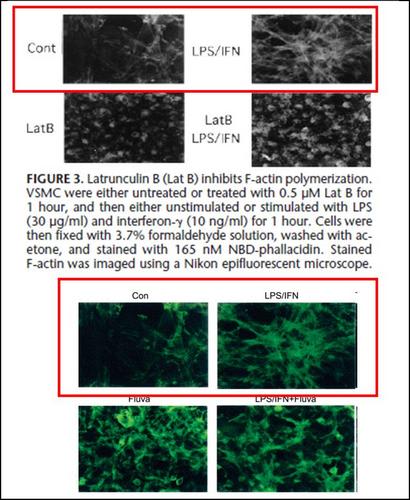
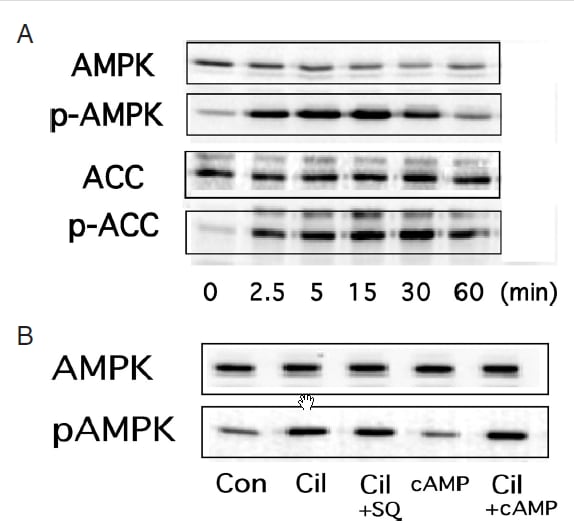
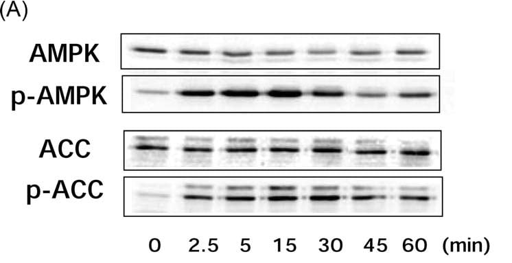
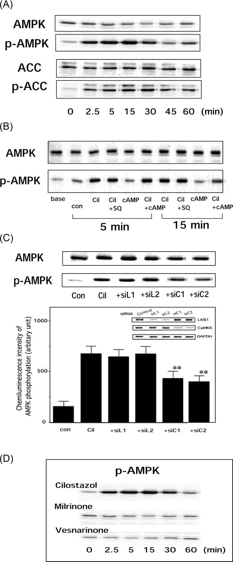
Fig.3 (A) AMPK activation by cilostazol. Cells were treated with different concentrations of cilostazol for 15 min and AMPK phosphorylation was assessed by Western blotting analysis.

論文#4

2011年01月24日 | 論文捏造
論文番号 著者名 論文タイトル名 発表雑誌名 巻、号 ページ 出版年
論文#4 Hattori Y, Suzuki K, Tomizawa A, Hirama N, Okayasu T, Hattori S, Satoh H, Akimoto K, Kasai K. Cilostazol inhibits cytokine-induced nuclear factor-kappaB activation via AMP-activated protein kinase activation in vascular endothelial cells. Cardiovasc Research 81(1) 133-139 2009


論文番号
指摘項目番号

不正が疑われる図の番号

論文#4 指摘項目No.1
指摘項目No.2
指摘項目No.3-a,b
指摘項目No.4
指摘項目No.5
指摘項目No.7
指摘項目No.8
指摘項目No.9
指摘項目No.10
指摘項目No.11
指摘項目No.19
指摘項目No.20
Figure 1(A),(D), 4(A) 
Figure 1(A),(D)
Figure 1(B)
Figure 1(A)
Figure 4(A)
Figure 4(B)
Figure 4(C)
Figure 4(C)
Figure 4(C)
Figure 4(C)
Figure 1(C)
Figure 1(C)


(A)- 指摘項目No.1
論文#4のFigure 4(A)の最上段のIkBαの画像の右3列分(左から4列目、5列目、6列目)の画像は、
同論文#4のFigure 1(A)のpAMPKの画像の左から2列目、3列目、4列目の画像と類似しています。
さらに、論文#4のFigure 4(A)の最上段のIkBαの画像(最上段の)の左から1列目、2列目の画像は、
同論文#4のFigure 1(A)のpAMPKの画像の左から5列目、6列目の画像や、
論文#9のFigure 1(a)のpAMPK画像の左から5列目、6列目の画像と、
類似しています(縦横比が改変されています。)
また、この論文#4のFigure1(A)のpAMPKの全7列分の画像は、
論文#9のFigure 1(a)のp-AMPKの全7列分の画像と類似しており、
また、これら#4,#9の二つの画像の左6列分と、
文#4のFigure 1(D)のpAMPKのCilostazolの画像全6列分や、
論文#3のFigure 2(A)のpAMPKの画像全6列分や、
論文#5のFigure 3(b)のMetformin pAMPKの画像全6列分とも
類似していることから、データの捏造(流用)が疑われます。
また、この論文#3のFigure 2(A)のpAMPKの画像の右5列分(2.5 min~60 minの画像)を、
上下左右逆にしたものが、同論文#3のFigure 3 (A)のpAMPKの画像に類似しています。
以上より、データの捏造(流用)が疑われます。


(A)- 指摘項目No.2
論文#4のFigure 1(A)のAMPKの画像の全7列分は、
論文#9のFigure 1(a)のAMPKの画像の全7列分と類似しています。
さらに、この論文#4と#9の画像のうち、左6列分(左から1,2,3,4,5,6列目)が、
論文#3のFigure 2(A)のAMPKの画像に類似しています。
さらに、この論文#3のFigure 2(A)のAMPKの画像6列分のうち、右5列分が、
同論文#3のFigure 3(A)のAMPKの画像に類似しています。
さらに、上記の論文#4と#9の画像のうち、右6列分(左から2,3,4,5,6,7列目)が、
論文#4のFigure 1(D)のpAMPKのMilrinoneの画像に類似しています(コントラストと縦横比を変更しています。)
以上より、データの捏造(流用)が疑われます。


(A)- 指摘項目No.3-a
論文#4のFigure 1(B)のAMPKの画像10列分のうち、右側5列分が、
論文#3のFigure 2(B)のAMPKの画像全5列分や、
論文#9のFigure 1(b)のAMPKの画像全5列分に、類似しています(縦横比が変更されてます。)。
(A)- 指摘項目No.3-b
同様に、
論文#4のFigure 1(B)のpAMPKの画像10列分のうち、左から2,3,4,5,6,列目の5列分が、
論文#3のFigure 2(B)のpAMPKの画像全5列分や、
論文#9のFigure 1(b)のpAMPKの画像全5列分に、
類似しており、データの捏造(流用)が疑われます(縦横比が変更されてます。)。


(A)- 指摘項目No.4
さらに、
論文#4のFigure 1(A)のACC画像と、pACC画像の全7列分は、
論文#9のFigure 1(a)のACC画像と、pACC画像 の全7列分と、類似しています。
さらに、これら#4, #9の画像の左側6列分の画像が、
論文#3のFigure 2(A)のACC画像と、p-ACC画像の全6列分と、類似しており、
データの捏造(流用)が疑われます。


(A)- 指摘項目No.5
論文#4のFigure 4(A)のPhospho-IkBαの画像と、
論文#8のFigure 5(A)のPhospho-IkB-αの画像と、
論文#14のFigure 3(A)のPhospho-IkBaの画像と、
論文#21のFigure 6(A)の上から2段目のPhospho IκB-αの画像の、
4つの画像は全て類似しており、データの捏造(流用)が疑われます。


(A)- 指摘項目No.7
論文#4のFigure 4(B)のIkBαの画像と、
論文#8のFigure 5(B)のIkB-αの画像と、
論文#14のFigure 3(B)のIkBaの画像と、
論文#21のFigure 6(A)のONOO- IκB-αの画像の左4列の画像の、
4つの画像は全て類似しており、データの捏造(流用)が疑われます


(A)- 指摘項目No.8
論文#2のFigure 4(C)の最上段のGST-IkB-αの画像は、
論文#4のFigure 4(C)の最上段GST-IkBαの画像や、
論文#8のFigure 5(C)の最上段のIkB-αの画像と、
類似しており、画像の流用(データの捏造)が疑われます。


(A)- 指摘項目No.9
論文#4のFigure 4(C)のIKKα/βの画像と、
論文#8のFigure 1(A)のACCの画像と、
同論文#8のFigure 5 (C)のIKKα/βの画像の3つの画像が、
全て類似しており、データの捏造(流用)が疑われます。


(A)- 指摘項目No.10
論文#4のFigure 4(C)の最下段のGST-IkBαの画像と、
論文#8のFgure 5(C)の最下段のIkB-αの画像が、
類似しており、データの捏造(流用)が疑われます。
さらに、この二つの画像は、
論文#2のFigure 1Aの上から5段目のAktの画像を左右逆にした画像や、
同論文#2のFigure 4Cの最下段のGST-IkB-αの画像を上下左右逆にした画像とも、
類似しており、画像の流用(データの捏造)が疑われます。


(A)- 指摘項目No.11
さらに、この論文#4のFigure 4(C)の最下段のGST-IkBαの画像(論文#8のFigure 5(C)の最下段のIkB-αの画像)と、
論文#8のFigure 1(A)の Aktの画像は、
縦・横の比率は異なりますが、形が類似しており、画像の流用(データの捏造)が疑われます。


(A)- 指摘項目No.19
論文#4のFigure 1(C)のAMPKの画像の左4列分(左から1、2、3、4列目)の画像は、
論文#7のFigure 2(A)のAMPKの画像の左4列分(左から1、2、3、4列目)の画像と
類似しており(縦横の比率が変更されています)、データの捏造(流用)が疑われます。 


(A)- 指摘項目No.20
論文#4のFigure 1(C)のpAMPKの画像の左3列分(左から1、2、3列目)の画像は、
論文#7のFigure 2(A)のpAMPKの画像の左3列分(左から1、2、3列目)の画像と
類似しており(縦横の比率が変更されています)、データの捏造(流用)が疑われます。


研究不正に関連する図(Figure)


↓論文#4のFigure 1(A)(画像クリックで解像度の高い画像が表示されます。)

Figure 1
(A) Cilostazol activates AMP-activated protein kinase (AMPK) in vascular endothelial cells. Human umbilical vein endothelial cells (HUVEC) were treated with cilostazol (100 μM) for the indicated time periods before lysis, after which each cell lysate sample was probed with antibodies specific for phosphorylated forms of AMPK and acetyl-CoA carboxylase (ACC).
Hattori Y et al. Cardiovasc Res 2009;81:133-139
Copyright the European Society of Cardiology.


↓論文#4のFigure 1(画像をクリックすると解像度の高い画像が表示されます。)

Copyright © 2011 European Society of Cardiology
Cardiovasc Res (2009) 81 (1): 133-139.
Figure 1
(A) Cilostazol activates AMP-activated protein kinase (AMPK) in vascular endothelial cells. Human umbilical vein endothelial cells (HUVEC) were treated with cilostazol (100 μM) for the indicated time periods before lysis, after which each cell lysate sample was probed with antibodies specific for phosphorylated forms of AMPK and acetyl-CoA carboxylase (ACC). (B) HUVEC were treated with cilostazol (100 μM) alone or in the presence of an adenylate cyclase inhibitor SQ 22536 (10 μM) or a cell-permeable cyclic AMP (cAMP) analogue pCTP-cAMP (100 μM). After 5 and 15 min of incubation, the cells were lysed and p-AMPK was analysed. Three independent studies showed similar results. (C) Cilostazol activates AMPK, which was significantly attenuated in HUVEC transfected with CaMKKβ siRNA (siC1 or siC2: 10 nM) but not with LKB1 siRNA (siL1 or siL2: 10 nM). Inset (lower figure): 48 h after cells were transfected with control siRNA, siL1, siL2, siC1, or siC2, the mRNA levels of LKB1, CaMKKβ, and GAPDH were determined. (D) Cilostazol, but not milrinone or vesnarinone, activates AMPK in vascular endothelial cells. HUVEC were treated with cilostazol (100 μM), milrinone (100 μM), or vesnarinone (100 μM) for the indicated time periods before lysis, after which each cell lysate sample was probed with antibodies specific for phosphorylated AMPK.


↓論文#4のFigure 4

論文#5

2011年01月24日 | 論文捏造
論文番号 著者名 論文タイトル名 発表雑誌名 巻、号 ページ 出版年
論文#5 Nakano A, Hattori Y, Aoki C, Jojima T, Kasai K. Telmisartan inhibits cytokine-induced nuclear factor-kappaB activation independently of the peroxisome proliferator-activated receptor-gamma. Hypertens Research 32(9) 765-769 2009


論文番号
指摘項目番号

不正が疑われる図の番号

論文#5 指摘項目No.1
Figure 3(b)


(A)- 指摘項目No.1
論文#4のFigure 4(A)の最上段のIkBαの画像の右3列分(左から4列目、5列目、6列目)の画像は、
同論文#4のFigure 1(A)のpAMPKの画像の左から2列目、3列目、4列目の画像と類似しています。
さらに、論文#4のFigure 4(A)の最上段のIkBαの画像(最上段の)の左から1列目、2列目の画像は、
同論文#4のFigure 1(A)のpAMPKの画像の左から5列目、6列目の画像や、
論文#9のFigure 1(a)のpAMPK画像の左から5列目、6列目の画像と、
類似しています(縦横比が改変されています。)
また、この論文#4のFigure1(A)のpAMPKの全7列分の画像は、
論文#9のFigure 1(a)のp-AMPKの全7列分の画像と類似しており、
また、これら#4,#9の二つの画像の左6列分と、
論文#4のFigure 1(D)のpAMPKのCilostazolの画像全6列分や、
論文#3のFigure 2(A)のpAMPKの画像全6列分や、
論文#5のFigure 3(b)のMetformin pAMPKの画像全6列分とも
類似していることから、データの捏造(流用)が疑われます。
また、この論文#3のFigure 2(A)のpAMPKの画像の右5列分(2.5 min~60 minの画像)を、
上下左右逆にしたものが、同論文#3のFigure 3 (A)のpAMPKの画像に類似しています。
以上より、データの捏造(流用)が疑われます。


研究不正に関連する図(Figure)


↓論文#5のFigure 3(B)

Figure 3 (b) Effect of the AMPK inhibitor compound C (CC) on
the telmisartan-mediated inhibition of TNFa-induced NF-kB activation.
Inset: Time course analysis for pAMPK by telmisartan or metformin. Data
represent the means±s.d. (n¼4). **Po0.01 compared with the control
value (TNFa only). NS, not significant between the absence and presence
of CC.
Copyright 2009 The Japanese Society of Hypertension
Hypertension Research (2009) 32, 765–769


論文#6

2011年01月24日 | 論文捏造
論文番号 著者名 論文タイトル名 発表雑誌名 巻、号 ページ 出版年
論文#6 Jojima T, Suzuki K, Hirama N, Uchida K, Hattori Y. Glimepiride upregulates eNOS activity and inhibits cytokine-induced NF-kappaB activation through a phosphoinoside 3-kinase-Akt-dependent pathway. Diabetes, Obesity and Metabolism 11(2) 143-149 2009
 
論文番号
指摘項目番号

不正が疑われる図の番号


論文#6 指摘項目No.33
指摘項目No.34
Figure 2(B)
Figure 2(B)


(A)- 指摘項目No.33
論文#2のFigure 4Aの最上段のIkB-αの左から2,3,4列目の3列分は、
同論文#2のFigure 4BのIkB-αの画像の右3列分(Fenofibrate 0, 30, 100 μMの画像)の画像と類似しています(画像サイズを加工しています)。
さらに、この3列分の画像は、
論文#6のFigure 2(B)の最上段のp-Aktの画像の左3列分(0, 5, 15 minの画像)と
類似しており、画像の流用(データの捏造)が疑われます。


(A)- 指摘項目No.34
論文#2のFigure 4BのIkB-αの画像の一番左の列の画像(Contの画像)は、
論文#6のFigure 2(B)の一番上のp-Aktの画像の左から4列目の画像(30 minの画像)と
類似しており、画像の流用(データの捏造)が疑われます。


研究不正に関連する図(Figure)

↓上記論文#6のFigure 2 


© 2008 The Authors
Journal Compilation © 2008 Blackwell Publishing Ltd
Diabetes, Obesity and Metabolism, 11, 2009, 143–149
Fig. 2 (A) Effect of two specific PI3K inhibitors, wortmannin
(WT) and LY294002 (LY), on glimepiride-induced
nitric oxide (NO) production. Human umbilical vein endothelial
cells (HUVEC) were preincubated with WT
(100 nmol/l) or LY294002 (20 mmol/l) for 30 min, after
which the cells were incubated with glimepiride (10 mmol/
l) for 60 min, and released NO was measured. Results represent
the mean  s.d. **p < 0.01 vs. glimepiride; n &amp;amp;#188; 3.
##p < 0.01 vs. basal; n &amp;amp;#188; 3. (B) Effect of glimepiride on
phosphorylation of Akt and endothelial NO synthase
(eNOS). HUVEC were incubated with glimepiride
(10 mmol/l) for the indicated times, and cell lysate samples
were prepared and subjected to Western blot analysis
using an antiphospho-Akt antibody and an antiphosphoeNOS
antibody.


論文#7

2011年01月24日 | 論文捏造
論文番号 著者名 論文タイトル名 発表雑誌名 巻、号 ページ 出版年
論文#7 Hattori Y, Nakano Y, Hattori S, Tomizawa A, Inukai K, Kasai K. High molecular weight adiponectin activates AMPK and suppresses cytokine-induced NF-κB activation in vascular endothelial cells FEBS Letters 582(12) 1719-1724 2008


論文番号
指摘項目番号

不正が疑われる図の番号


論文#7 指摘項目No.19
指摘項目No.20
Figure 2(A)
Figure 2(A)


研究不正に関連する図(Figure)

↓論文#7のFigure 2(画像をクリックすると解像度の高い画像が表示されます。)

Copyright © 2008 Published by Elsevier B.V.
FEBS Letters Volume 582, Issue 12, 28 May 2008, Pages 1719-1724
Fig. 2. (A) HMW adiponectin (Ad) activates AMPK, which was significantly attenuated in HUVEC transfected with CaMKKβ siRNA (siC: 10 nM) but not with LKB1 siRNA (siL: 10 nM). Inset (lower figure): 48 h after cells were transfected with control, LKB1, or CaMKKβ siRNA, the mRNA levels of LKB1 or CaMKKβ were determined. (B) HMW adiponectin (Ad)-induced AMPK phosphorylation was inhibited by the inhibitor of CaMKKβ, STO-609 in HUVEC. Results represent the means ± S.D. (n = 4). **P < 0.01 vs. AMPK activity by Ad.


論文#8

2011年01月24日 | 論文捏造
論文番号 著者名 論文タイトル名 発表雑誌名 巻、号 ページ 出版年
論文#8 Okayasu T, Tomizawa A, Suzuki K, Manaka K, Hattori Y. PPARalpha activators upregulate eNOS activity and inhibit cytokine-induced NF-kappaB activation through AMP-activated protein kinase activation. Life Sciences 82(15-16) 84-891 2008


論文番号
指摘項目番号

不正が疑われる図の番号


論文#8 指摘項目No.5
指摘項目No.7
指摘項目No.8
指摘項目No.9
指摘項目No.10
指摘項目No.11
指摘項目No.12
指摘項目No.13
指摘項目No.29
指摘項目No.30
指摘項目No.31
Figure 5(A)
Figure 5(B)
Figure 5(C)
Figure 1(A)
Figure 5(C)
Figure 1(A), 5(C)
Figure 1(A),(B)
Figure 5(A)
Figure 1(A)
Figure 1(A)
Figure 1(A)


(A)- 指摘項目No.5
論文#4のFigure 4(A)のPhospho-IkBαの画像と、
論文#8のFigure 5(A)のPhospho-IkB-αの画像と、
論文#14のFigure 3(A)のPhospho-IkBaの画像と、
論文#21のFigure 6(A)の上から2段目のPhospho IκB-αの画像の、
4つの画像は全て類似しており、データの捏造(流用)が疑われます。


(A)- 指摘項目No.7
論文#4のFigure 4(B)のIkBαの画像と、
論文#8のFigure 5(B)のIkB-αの画像と、
論文#14のFigure 3(B)のIkBaの画像と、
論文#21のFigure 6(A)のONOO- IκB-αの画像の左4列の画像の、
4つの画像は全て類似しており、データの捏造(流用)が疑われます


(A)- 指摘項目No.8
論文#2のFigure 4(C)の最上段のGST-IkB-αの画像は、
論文#4のFigure 4(C)の最上段GST-IkBαの画像や、
論文#8のFigure 5(C)の最上段のIkB-αの画像と、
類似しており、画像の流用(データの捏造)が疑われます。


(A)- 指摘項目No.9
論文#4のFigure 4(C)のIKKα/βの画像と、
論文#8のFigure 1(A)のACCの画像と、
同論文#8のFigure 5 (C)のIKKα/βの画像の3つの画像が、
全て類似しており、データの捏造(流用)が疑われます。


(A)- 指摘項目No.10
論文#4のFigure 4(C)の最下段のGST-IkBαの画像と、
論文#8のFgure 5(C)の最下段のIkB-αの画像が、
類似しており、データの捏造(流用)が疑われます。
さらに、この二つの画像は、
論文#2のFigure 1Aの上から5段目のAktの画像を左右逆にした画像や、
同論文#2のFigure 4Cの最下段のGST-IkB-αの画像を上下左右逆にした画像とも、
類似しており、画像の流用(データの捏造)が疑われます。


(A)- 指摘項目No.11
さらに、この論文#4のFigure 4(C)の最下段のGST-IkBαの画像(論文#8のFigure 5(C)の最下段のIkB-αの画像)と、
論文#8のFigure 1(A)の Aktの画像は、
縦・横の比率は異なりますが、形が類似しており、画像の流用(データの捏造)が疑われます。


(A)- 指摘項目No.12
論文#8のFigure 1 (A)のp-AKTの画像と、
同論文のFigure 1(B)のp-ACCの画像が、
類似しており、画像の流用(データ捏造)が疑われます。


(A)- 指摘項目No.13
論文#8のFigure 5(A)のIkB-αの画像と、
論文#14のFigure 3(A)のIkB-aの画像と、
論文#21のFigure 6(A)のIkB-αの画像とが、
類似しており、画像の流用(データ捏造)が疑われます。


(A)- 指摘項目No.29
論文#2のFigure 1Aの上から1段目と2段目のAMPKとpAMPKの画像は、
論文#8のFigure 1(A)の上から1段目と2段目のAMPKとpAMPKの画像と、
類似しており、画像の流用(データの捏造)が疑われます。


(A)- 指摘項目No.30
論文#2のFigure 1Aの上から6段目のp-Aktの画像は、
論文#8のFigure 1(A)の上から6段目のp-Aktの画像と、
類似しており、画像の流用(データの捏造)が疑われます。


(A)- 指摘項目No.31
論文#2のFigure 1Aの上から7段目のeNOSの画像は、
論文#8のFigure 1(A)の上から7段目のeNOSの画像を左右逆に加工した画像と同一であり、
画像の流用(データの捏造)が疑われます。


研究不正に関連する図(Figure)

↓論文#8のFigure 1(A)(B)


© 2008 Elsevier Inc. All rights reserved.
Life Sciences 82 (2008) 884–891
Fig. 1. (A) Fenofibrate activates AMPK in vascular endothelial cells. HUVEC were treated with 100 μmol/L fenofibrate for the indicated time periods before lysis, after which cell lysates were probed with antibodies specific for AMPK, ACC, Akt, or eNOS, or their phosphorylated forms. (B) WY14643 (100 μmol/L) activates AMPK in HUVEC.


↓論文#8のFigure 5


© 2008 Elsevier Inc. All rights reserved.
Life Sciences 82 (2008) 884–891
Fig. 5. (A) HUVEC were incubated with TNFα for 0–180 min. The cells were lysed and subjected to Western blot analysis using anti-IκB-α and anti-phospho-IκB-α antibodies. (B) The effect of fenofibrate on IκB-α degradation in HUVEC. Cells were incubated for 30 min with fenofibrate (30 and 100 μM), followed by TNFα for 30 min. Cells were then lysed and subjected to Western blot analysis using anti-IκB-α antibody. (C) The effect of fenofibrate on IKK activity in HUVEC. Cells were incubated for 30 min with metformin (30 and 100 μM), followed by TNFα for 15 min. Cells were then lysed and immunoprecipitated with anti-IKKα/β antibody and used for kinase assay using recombinant IκB-α as a substrate. Note that equal band densities for IKKα/β- and IκB-α were observed. Three independent experiments for (A) and (B) and two independent experiments for (C) were performed. Similar results were obtained in each experiment and representative photos were shown.