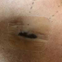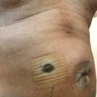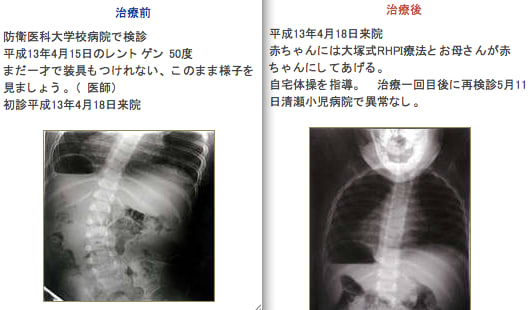
(添付写真は大塚整体ホームページより引用:)
august03より:側彎症は正しい診断に基づく正しい治療が必要です。
正しい医学を学び、適切な診断機器を持つ専門医師のいる医療機関でのみ治療が
可能です。たまたま幸運にも、障害を有しない赤ちゃんだったからよかったものの
一歩間違えたら、この赤ちゃんは死亡する危険性さえあったでしょう。
医師(medical doctor)でもない者が、このような宣伝を繰り広げることは患者の
利益に反する社会悪以外のなにものでもないと私augsut03は考えます。
“脊柱が曲がるという病態”を [側弯症] という言葉でひとくくりにしてイメージして
いますが、原因が不明である[特発性側弯症]は別格としても、原因が明らかにされ
いる側弯症もあることを、皆さんは知識として持っておいていただきたいと思い
ます。
側弯症と診断される患者さんの大多数が[思春期の女の子に多い特発性側弯症]で
あり、その原因が不明であることから、全ての側弯症が、原因不明の病気という
間違った認識が持たれている傾向もあります。
ここでは原因を特定している文献をアブストラクトのみですが、ご紹介したいと
思います。
なお、この中の中心的医学用語 Neural Axis Abnormalities の正確な和訳が
見つからないため、下記では「神経軸障害」という言葉を当てはめました。
側彎症の種類のひとつに「症候性側弯」がありますが、この「神経軸障害」
も、症候性側弯のように、生来的に脊髄/神経に障害があり、それが原因で脊柱が
曲がってくる、という機序というのが私の理解です。もし、私の理解不足がござい
ましたら、ご指導いただければと思います。
(参考:症候性側弯 http://www7a.biglobe.ne.jp/~orthopedics/scoliosissynd.htm)
(ブログ内の参照)
「SRS BRACE MANNUAL 側わん症装具マニュアル No.3」
http://blog.goo.ne.jp/august03/e/e749759d03bfa433bc1d082ac4d90685
脊髄空洞症は正確には [syringomyelia]が英語での専門用語になりますので、
この文献上で記載されている neuro-axis defectsはさらに大きな範疇としての
神経(脊髄)系の疾患として用いられているようです。
語の成り立ちは、 neuro /神経 axis/軸 defects/欠損、であり、また
defectに代えて、neural axis abnormalitiesという表記をしている文献もあり、
神経系(脊髄)になんらかの異常があることを示しています。それらには
syringomyelia(脊髄空洞症), Arnold Chiari malformations(キアリ奇形),
tethered cords(二分脊椎), spinal cord tumor(脊髄腫瘍) などが含まれている
ようです。
------------------------------------------------------------------------
(文献1)
Incidence of Neural Axis Abnormalities in Infantile Patients Diagnosed
with Idiopathic Scoliosis: Is a Screening MRI Necessary?
Abstract from the SRS 2001 Annual Meeting
特発性側弯症と診断された乳幼児期側弯症における神経軸障害の発生率:
MRI検査は必須であるか ?
http://www.spineuniverse.com/displayarticle.php/article1473.html
Lawrence G. Lenke, M.D.*
Washington University School of Medicine, St. Louis, MO, USA,
米国ミズリー州セントルイス ワシントン大学
はじめに:文献によれば、20度を超えるカーブの学童期特発性側わん症患者のうち
約20%に神経軸障害があることが報告されている。乳幼児期特発性側彎症に
どれくらい神経軸障害があるかについてはよく調査されていなかった。
これらの若年のこども達に対してMRIでのスクリーニング調査をするかどうかの決定
には鎮静剤の必要性や全身麻酔の実施などを考慮しなければならない。
目的:大規模で、連続性を保った多施設医療機関での乳幼児期特発性側彎症患者に
おける神経軸障害の有無を調査すること。また、この年代のこどもたちに
対してMRIスクリーニングの必要性を調査すること。
方法:多施設によるレトロスペクティブ(後ろ向き研究)レビューにより56人を評価
した。期間は1993~2000年、脊椎専門3医療機関で、次の基準にあう患者とした。
特発性側わん症と診断された / 3歳以下 / 20度以上のカーブ / 神経症状はない
/ 症候性側彎ではない / 先天性障害はない。
全ての患者が頭蓋から尾骨まで(仮訳)神経索障害を調査するためにMRIで検査した。
結果:56人のうち11人(19.6%)の患者に神経軸障害が見つかった。この内訳は
脊髄空洞症5人、アーノルドキアリ奇形3人、係留脊髄症2人、脊髄腫瘍1人であった。
神経軸障害を有する11のうち、6人はその障害に対して神経学的手術を必要
としていた。
考察:これまでに行われたふたつの研究では、わずか10人の乳幼児期特発性側わん症
が報告され、そのうち5人(50%)に神経軸障害があった。今回の大規模な調査
結果から、現実的には、約20%に神経軸障害があると予想される。
結論: 乳幼児期特発性側わん症患者の19.6%に神経軸障害が見つかった。
これは、これまで報告されていた学童期特発性側わん症患者での神経軸障害
の発見率とほぼ同じであった。この高い発症率と、この障害を有する乳幼児患者の
54.5%では神経外科手術を要していたということから、20度以上のカーブを有する
乳幼児側彎症患者には、脊椎全体に対するMRI検査が推奨される
(文献2)
Neural Axis Abnormalities in Congenital Spine Deformity:
Is a Screening MRI Necessary?
先天性の脊椎障害における神経軸障害:MRI検査は必要か ?
http://www.spineuniverse.com/displayarticle.php/article2299.html
目的:MRI検査をルーティンに行うことの価値は最近の文献においてずっと議論
されてきている話題である。本研究は、障害の発生状況、脊椎奇形のタイプ、
神経学的検証やそれらの奇形の特定としてのレントゲン評価などを目的とする。
方法:ひとつの医療機関において、1992年~1999年に、先天性脊椎奇形を有する
97人(59女子、38男子)の患者をMRIで検査した。すべての診療記録、レントゲン、
MRI検査結果をレビューした。57人は先天性側わん症、27人は先天性前わん側わん症
、13人は先天性前わん症であった。平均年齢は4歳±3歳(1ヶ月~13歳)。
結果:97人のうち24人(24.7%)に、MRI検査で脊柱管内に奇形が見つかった。
(27.1%女子、21.1%男子)。それらの奇形の内訳は、脊髄正中離(diastematomyelia)14人、
脊髄係留症(tethered cord)11人、脊髄空洞症(syringomyelia)4人、Dural Ectasia4人、
Lipoma1人。8人には複合した奇形が見られた。
MRIで異常を発見した24人のうち、16人はレントゲン検査では異常は見つけられず、
18人では神経症状検査では正常であった。脊柱管内奇形を発見するうえでの診断に
よる正確度は25%、レントゲン写真による正確度は33.4%であった。奇形は、腰椎に
もっとも多く見られ(9人)、unilateral barsに関係していた。8人は外科手術を必要
とした。
結論: 神経軸障害を発見する率は、診断やレントゲン写真では低いものとなる。
先天性脊椎奇形を有する患者においては、MRI検査は必須の検査である。
三分の一の患者で手術を必要とした、ということからも、MRIによる早期の検査を
実施することは、神経障害のさらなる悪化を防ぐ意味からも重要である。
--------------------------------------------------------------------
(備考)
脊髄空洞症に対する手術はあくまでも症状の進行を予防するものであり、
病状が進行してから手術を行っても、運動麻痺や知覚障害の回復はあまり期待
できません。早期に診断・治療を受けることが重要です。
/////////////////////////////////////////////////////////////////////
ブログ内の関連記事
「側弯症と脊髄空洞症」
http://blog.goo.ne.jp/august03/e/c0dbc1945c1e9668ffabb7599150cacd
「医行為および医師法17条について」
http://blog.goo.ne.jp/august03/e/ae1f9df6503e61deced6f883ead765d7
こどもたちの背中を整体から守るキャンペーン
~ 私たちは患者を保護するために整体を規制する法制化を求めます ~
もし趣旨にご賛同いただけましたら、上記の言葉をネット上でご使用いただけます
よう、ご協力を御願いします。
(2008年6月7日追記)
Neural Axis Abnormalitiesの訳について、「整形外科医のひとりごと」の
Skelton先生よりアドバイスをいただきました。
言葉としての「neural axis」は「神経軸」で良いかと思いますが。具体的には online medical dictionary からの引用ですが、
neural axis → cerebrospinal axis =The central nervous system;
the brain and spinal cord. Synonym: encephalomyelonic axis, neural axis
とあります。
つまり「中枢神経系」ということになりますが、あえてこうした言葉を使うのは脳だけの障害ではなく、脊髄の障害を主眼にして発生学的な面からも考えているからかもしれませんね。
(文献1)
INTRODUCTION: Although the literature demonstrates approximately 20%
incidence of neural axis abnormalities in juvenile idiopathic scoliosis
patients with curves >20º, the incidence of neural axis abnormalities in
the infantile idiopathic population is not well documented. The decision to
obtain a screening MRI on these young children is also tempered with the
necessity for IV sedation or even a general anesthetic to obtain the study.
PURPOSE: To determine the incidence of neural axis abnormalities in
infantile idiopathic scoliosis patients without neurologic findings in a
large, consecutive, multi-center series, to determine the need for a
screening MRI in this age group.
METHODS: A multi-centered retrospective review evaluated 56 consecutive
patients between 1993-2000 at 3 spinal deformity clinics with the following
inclusion criteria: presumed idiopathic scoliosis at presentation; age 3 or
less; scoliosis > 20º; no neurological findings; no associated syndromes;
no congenital abnormalities. All patients were evaluated by a total spine
MRI protocol for examination of neural axis abnormalities from the skull to
the coccyx.
RESULTS: 11 of 56 (19.6%) of the patients were found to have a neural axis
abnormality on their MRI. This included 5 with syringomyelia, 3 with Arnold
Chiari malformations, 2 with tethered cords, and one with a spinal cord
tumor. Of these 11 patients with abnormalities, 6 needed neurosurgical
intervention for their abnormality for an incidence of 54.5%.
。
DISCUSSION: In two previous studies with a combined total of only 10
infantile idiopathic patients, 5 (50%) were noted to have a neural axis
abnormality on MRI evaluation. The actual incidence appears to be closer to
20% in this significantly larger multi-center study.
CONCLUSIONS: The 19.6% incidence of neural axis abnormalities in the
infantile idiopathic scoliosis group was found to be almost identical to
that reported in the literature for the juvenile idiopathic patients. Due
to this high incidence and the fact that 54.5% of the infantile patients
with abnormal MRI's required neurosurgical intervention, a total spine MRI
is recommended at presentation in patients with infantile idiopathic
scoliosis >20º.
(文献2)
Purpose: The value of obtaining routine MRI study has been debated in
recent literature. This study was undertaken to identify the incidence,
type of intraspinal anomaly and validity of neurological (N/E) and
radiographic evaluation in detecting these anomalies.
Methods: Ninety-seven patients (59 female, 38 male) with congenital spinal
deformity seen in one institution between 1992 and 1999 who had screening
MRI were studied. All medical records, plain radiographs and MRI studies
were reviewed. Fiftyseven had congenital scoliosis, 27 congenital
kyphoscoliosis and 13 congenital kyphosis. The mean age at presentation was
4+3 years (1 mo to 13 years)
Results: Twenty-four of 97 (24.7%) of the patients were found to have
intraspinal anomalies on their MRI (27.1% of females and 21.1% of males).
These anomalies included: Diastematomyelia (14), Tethered cord (11),
Syringomyelia (4), Dural Ectasia (4) and Lipoma (1). There were 8 complex
anomalies. Of 97 patients, 20 had abnormal neurological examination and 10
had abnormal radiographs.
Of 24 patients with abnormal MRI, 16 had normal radiographs and 18 had
normal neurological examination. The sensitivity of clinical and
radiographic assessment in detecting intraspinal anomalies was 25% and
33.4% respectively. The specificity of assessments was also 81% and 97.3%.
Anomalies were more frequent in lumbar spine (9) and mostly associated with
unilateral bars. Eight patients (1/3) required surgical treatment for their
underlying anomalies.
Conclusions: The yield for detecting neural axis anomalies based on
clinical and radiographic evaluation is low. MRI is an integral part of the
assessment of patients with congenital spinal deformity. Since 1/3 of
patients require surgical intervention, it should be done early in the
course of treatment to prevent further neurological deficit.