


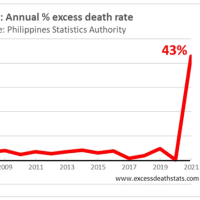
5 Christopher Exley博士 自閉症の脳組織中のアルミニウム JTEMB 2018
Fig. 4. Intracellular aluminium in cells morphologically compatible with microglia within the parietal and temporal lobes of 29-year-old (A8) and 15-year-old (A4) male donors, diagnosed with autism.
図 4. 自閉症と診断された 29 歳 (A8) および 15 歳 (A4) の男性ドナーの頭頂葉および側頭葉内のミクログリアと形態学的に適合する細胞の細胞内アルミニウム。
Lumogallion-reactive extracellular aluminium (white arrows) producing an orange fluorescence emission was noted around likely microglial cells in the parietal (a) and temporal lobes (b) of donors A8 and A4 respectively.
オレンジ色の蛍光発光を生成するルモガリオン反応性細胞外アルミニウム(白い矢印)は、それぞれドナーA8およびA4の頭頂葉( a )および側頭葉( b )のミクログリア細胞の周囲に認められました。
Non-stained adjacent (5 μm) serial sections, produced a weak green autofluorescence emission of the identical area imaged in white (c) and grey matter (d) of the respective lobes.
染色されていない隣接する(5μm)連続切片は、それぞれの葉の白( c )および灰白質( d )で画像化された同一領域の弱い緑色の自己蛍光発光を生成しました。
Upper and lower panels depict magnified inserts marked by asterisks, of the fluorescence channel and bright field overlay.
上部と下部のパネルは、蛍光チャネルと明視野オーバーレイの、アスタリスクでマークされた拡大された挿入を示しています。
Magnification ×400, scale bars: 50 μm.
倍率×400、スケールバー:50μm。
(For interpretation of the references to colour in this figure legend, the reader is referred to the web version of this article.)
(この図の凡例における色への言及の解釈については、読者はこの記事の Web バージョンを参照してください。)


